

Hematologic Malignancies of the Breast: A Contemporary Series Investigating Incidence, Presentation, Accuracy of Diagnosis on Core Needle Biopsy, and Hormone Receptor Expression
- PMID: 30814841
- PMCID: PMC6385330
- DOI: 10.1177/1178223419830982
Hematologic Malignancies of the Breast: A Contemporary Series Investigating Incidence, Presentation, Accuracy of Diagnosis on Core Needle Biopsy, and Hormone Receptor Expression
Abstract
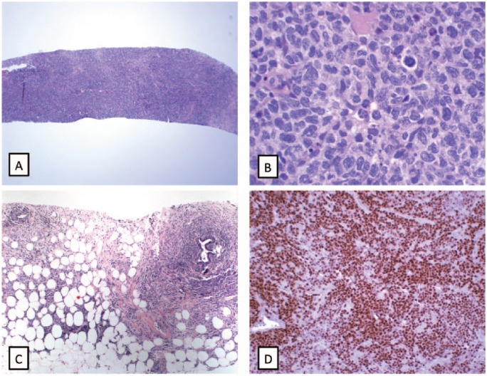
Background: Distinguishing breast hematologic malignancies in core needle biopsies from other entities can be challenging. Misclassification as a breast carcinoma could result in inappropriate treatment. The aim of this study was to characterize the types, incidence, and helpful diagnostic features of hematologic malignancies of the breast.
Design: All hematologic malignancies of the breast diagnosed at our institution from 2004 to 2017 were identified. Clinical notes, imaging, and slides were reviewed. Immunohistochemical analysis of estrogen receptor α (ERα), estrogen receptor β (ERβ), and androgen receptor (AR) was performed when tissue was available.
Results: In all, 43 hematologic malignancies from biopsies of 37 women and 6 men were identified. Core needle biopsies (35 or 81%) were more common than excisions (8 or 19%). For 14 patients (40%), the core biopsy was the first diagnosis of a hematologic malignancy. Diagnoses included 37 lymphomas (7 primary), 4 leukemias, and 2 myelomas. There was 1 misdiagnosis of carcinoma. Low positivity for hormone receptors was observed in a minority of lymphomas. A definitive diagnosis of hematologic malignancy was made in 31 (89%) of the core needle biopsies. Only 3 patients undergoing core biopsy required excision for diagnosis.
Conclusions: Most of the hematologic malignancies of the breast are currently diagnosed on core needle biopsy and 40% of patients do not have a prior history. To avoid errors, pathologists need to be aware of diagnostic features and morphologic mimics. A hematologic malignancy should be considered if tumor cells are discohesive, carcinoma in situ is absent, and hormone expression is low or absent.
Keywords: breast hematologic malignancies; breast lymphoma.
Conflict of interest statement
Declaration of conflicting interests:The author(s) declared no potential conflicts of interest with respect to the research, authorship, and/or publication of this article.
Figures


Similar articles
-
Reflex Estrogen Receptor (ER) and Progesterone Receptor (PR) Analysis of Ductal Carcinoma In Situ (DCIS) in Breast Needle Core Biopsy Specimens: An Unnecessary Exercise That Costs the United States $35 Million/y.Am J Surg Pathol. 2016 Aug;40(8):1090-9. doi: 10.1097/PAS.0000000000000674. Am J Surg Pathol. 2016. PMID: 27299796
-
Office-based core needle biopsy of bone and soft tissue malignancies: an accurate alternative to open biopsy with infrequent complications.Clin Orthop Relat Res. 2010 Oct;468(10):2774-80. doi: 10.1007/s11999-010-1422-5. Epub 2010 Jun 26. Clin Orthop Relat Res. 2010. PMID: 20582496 Free PMC article.
-
Long term clinical follow-up of atypical ductal hyperplasia and lobular carcinoma in situ in breast core needle biopsies.Pathology. 2016 Jan;48(1):25-9. doi: 10.1016/j.pathol.2015.11.015. Epub 2015 Dec 14. Pathology. 2016. PMID: 27020205
-
Hematologic malignancies of the liver: spectrum of disease.Radiographics. 2015 Jan-Feb;35(1):71-86. doi: 10.1148/rg.351130008. Radiographics. 2015. PMID: 25590389 Review.
-
Prostate needle biopsies containing prostatic intraepithelial neoplasia or atypical foci suspicious for carcinoma: implications for patient care.J Urol. 2006 Mar;175(3 Pt 1):820-34. doi: 10.1016/S0022-5347(05)00337-X. J Urol. 2006. PMID: 16469560 Review.
Cited by
-
Metastatic cyclin D1 positive lobular breast carcinoma as a potential lymphoma mimicker.EJHaem. 2023 Sep 4;4(4):1174-1175. doi: 10.1002/jha2.781. eCollection 2023 Nov. EJHaem. 2023. PMID: 38024639 Free PMC article. No abstract available.
-
Primary breast lymphoma initially diagnosed as invasive ductal carcinoma: A case report.Clin Case Rep. 2021 May 5;9(6):e04189. doi: 10.1002/ccr3.4189. eCollection 2021 Jun. Clin Case Rep. 2021. PMID: 34194774 Free PMC article.
References
-
- Domchek SM, Hecht JL, Fleming MD, Pinkus GS, Canellos GP. Lymphomas of the breast: primary and secondary involvement. Cancer. 2002;94:6–13. - PubMed
-
- Aviv A, Tadmor T, Polliack A. Primary diffuse large B-cell lymphoma of the breast: looking at pathogenesis, clinical issues and therapeutic options. Ann Oncol. 2013;24:2236–2244. - PubMed
-
- Ishikawa MK, Pinsky RW, Smith LB, Jorns JM. Morphologic mimics of invasive lobular carcinoma. Arch Pathol Lab Med. 2015;139:1253–1257. - PubMed
-
- Hugh JC, Jackson FI, Hanson J, Poppema S. Primary breast lymphoma. An immunohistologic study of 20 new cases. Cancer. 1990;66:2602–2611. - PubMed
-
- Bobrow LG, Richards MA, Happerfield LC, et al. Breast lymphomas: a clinicopathologic review. Hum Pathol. 1993;24:274–278. - PubMed
LinkOut - more resources
Full Text Sources
Research Materials
