. 2018 Feb 15;42(1):63-75.
doi: 10.3906/biy-1706-6. eCollection 2018.

Ultrasound-enhanced gene delivery to alfalfa cells by hPAMAM dendrimer nanoparticles

Affiliations

Ultrasound-enhanced gene delivery to alfalfa cells by hPAMAM dendrimer nanoparticles

Amin Amani et al. Turk J Biol. .

Abstract

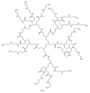
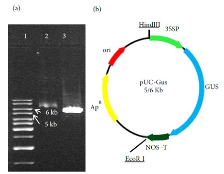
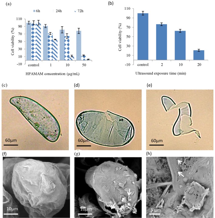
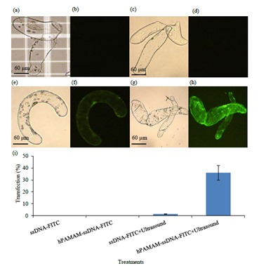
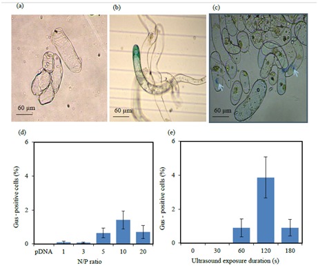
Cationic polyamidoamine (PAMAM) dendrimers are highly branched nanoparticles with unique molecular properties, which make them promising nanocarriers for gene delivery into cells. This research evaluated the ability of hyperbranched PAMAM (hPAMAM)-G2 with a diethylenetriamine core to interact with DNA, its protection from ultrasonic damage, and delivery to alfalfa cells. Additionally, the effects of ultrasound on the efficacy of hPAMAM-G2 for the delivery and expression of the gus A gene in the alfalfa cells were investigated. The electrophoresis retardation of plasmid DNA occurred at an N/P ratio (where N is the number of hPAMAM nitrogen atoms and P is the number of DNA phosphorus atoms) of 3 and above, and hPAMAM-G2 dendrimers completely immobilized the DNA at an N/P ratio of 4. The analysis of the DNA dissociated from the dendriplexes revealed a partial protection of the DNA from ultrasound damage at N/P ratios lower than 2, and with increasing N/P ratios, the DNA was better protected. Sonication of the alfalfa cells in the presence of ssDNA-FITC-hPAMAM increased the ssDNA delivery efficiency to 36%, which was significantly higher than that of ssDNA-FITC-hPAMAM without sonication. Additionally, the efficiency of transfection and the expression of the gus A gene were dependent on the N/P ratio and the highest efficiency (1.4%) was achieved at an N/P ratio of 10. The combination of 120 s of ultrasound and hPAMAM-DNA increased the gusA gene transfection and expression to 3.86%.

Keywords: Gene transfer; Medicago sativa L; hPAMAM-DNA complex; polyamidoamine dendrimers; sonication.


Figures

Figure 1
Figure 1
Schematic structure of hPAMAM dendrimers G1 with diethylenetriamine core.
Figure 2
Figure 2
The structure of pUC-gusA vector: (a) pUC-gusA recombinant vector digested with EcoRI, lane 1: 1-kb ladder, lane 2: uncut pUC-gusA recombinant vector, lane 3: pUC-gusA plasmid digested with EcoRI. (b) The schematic representation of pUC-gusA vector carrying gusA gene under control of the CaMV 35S promoter and NOS terminator.
Figure 3
Figure 3
Characteristics of hPAMAM–DNA complexes: (a) DNA mobility retardation assay of hPAMAM–DNA complexes at different N/P ratios, lane 1: N/P ratio of 0 (pDNA only), lanes 2–10: N/P ratios of 0.25, 0.5, 1, 2, 3, 4, 5, 10, and 20, respectively. (b) pDNA protection from endonuclease activity, pDNA dissociated from undigested (lane 1) and digested hPAMAM–DNA complex at N/P ratios of 0, 10, 20, 30, 40, and 50 (lanes 2–7, respectively). (c) pDNA protection from ultrasound damage: pDNA dissociated from nonsonicated (lane 1) and sonicated hPAMAM–DNA complex at N/P ratios of 0, 0.1, 0.2, 0.4, 0.6, 0.8, 1, and 2 (lanes 2–9, respectively). (d) SEM micrograph of G2 hPAMAM–DNA complex at N/P ratio of 10.
Figure 4
Figure 4
(a) The alfalfa cells’ viability under the effect of different concentrations and exposure durations of G2 hPAMAM; (b) the effect of different exposure durations of ultrasound on the alfalfa cells’ viability. The control (c) and sonicated (d and e) alfalfa cells were observed by light microscopy. The control cells (f) and changes caused by sonication (g and h) in alfalfa cells were analyzed by SEM.
Figure 5
Figure 5
Fluorescence microscopy analysis of the alfalfa cells. Bright-field (a) and fluorescence (b) images of the untreated (control) alfalfa cells. Bright-field (c) and fluorescence (d) images of the alfalfa cells incubated with ssDNA-FITC. Bright-field (e) and fluorescence (f) images of the alfalfa cells incubated with ssDNA-FITC and ultrasound. Bright-field (g) and fluorescence (h) images of the alfalfa cells incubated with hPAMAM-ssDNA-FITC and ultrasound. (i) Effects of hPAMAM dendrimers and ultrasound on the transfection of the alfalfa cells with ssDNA-FITC.
Figure 6
Figure 6
GUS histochemical assay. (a) Untreated (control) alfalfa cells and (b and c) the alfalfa cells transfected by hPAMAM–DNA complexes. The effects of different N/P ratios (d) and different durations of ultrasound exposure (e) on the efficiency of alfalfa cell transfection by hPAMAM–DNA complexes.

References

    1. Abdelhady HG , Lin YL , Sun H , ElSayed ME ( 2013. ). Visualizing the attack of RNase enzymes on dendriplexes and naked RNA using atomic force microscopy . PLoS One 8 : e61710 . - PMC - PubMed
    1. Al-Dosari MS , Gao X ( 2009. ). Nonviral gene delivery: principle, limitations, and recent progress . AAPS J 11 : 671 - 681 . - PMC - PubMed
    1. Altpeter F , Baisakh N , Beachy R , Bock R , Capell T , Christou P , Daniell H , Datta K , Datta S , Dix PJ et al. ( 2005. ). Particle bombardment and the genetic enhancement of crops: myths and realities . Mol Breeding 15 : 305 - 327 .
    1. Bande F , Arshad SS , Bejo MH , Kamba SA , Omar AR ( 2015. ). Synthesis and characterization of chitosan-saponin nanoparticle for application in plasmid DNA delivery . J Nanomater 16 : 207 .
    1. Bielinska AU , Kukowska-Latallo JF , Baker JR ( 1997. ). The interaction of plasmid DNA with polyamidoamine dendrimers: mechanism of complex formation and analysis of alterations induced in nuclease sensitivity and transcriptional activity of the complexed DNA . Biochim Biophys Acta 1353 : 180 - 190 . - PubMed

LinkOut - more resources