. 2019 Feb 13:13:11.
doi: 10.3389/fnana.2019.00011. eCollection 2019.

Berengario da Carpi and the Renaissance of Brain Anatomy

Affiliations

Berengario da Carpi and the Renaissance of Brain Anatomy

André Parent. Front Neuroanat. .

Abstract

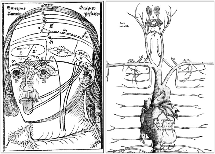
Berengario da Carpi (Jacopo Barigazzi) was born around 1460 in the small Italian town of Carpi near Modena. Berengario's father, Faustino, was a reputable barber-surgeon who initiated his son early into the art of anatomy and surgery. After his graduation from the University of Bologna in 1489, Berengario rapidly acquired an enviable reputation as a physician and surgeon following the successful treatment of several dignitaries, including Lorenzo de' Medici, Duke of Urbino who suffered a severe head injury in 1517. While professor of anatomy and surgery at the University of Bologna, Berengario published in 1518 his De fractura cranei, a landmark work on cranio-cerebral surgery. Berengario's masterpiece, however, is undoubtedly his detailed Commentaria on the famous medieval anatomy treatise of Mondino de' Liuzzi (ca. 1270-1326) that he published in 1521. A shorter version entitled Isagogae Breves appeared a year later. Besides a facsimile of Mondino's work, Berengario's Commentaria contains a wealth of new information, including observations that challenged Galenic physiology. Galen taught that the rete mirabile-a vascular plexus believed to occur at the basis of the human brain-is the locus where the vital spirit is transformed into the more sophisticated animal spirit that is stored in the brain ventricles to be later released at the periphery through a journey within hollow nerves. Courageously, Berengario wrote that despite many attempts he was unable to detect the famous rete mirabile in humans. He also noted that the nerves linked to the brain are solid structures, not hollow tubes, as advocated by Galen. His conclusions were based on a systematic dissection method that he called anatomia sensibilis, a term that emphasizes the sensory over textual versions of the truth. Berengario contributed significantly to human brain anatomy, with a detailed description of the meninges and cranial nerves and the first comprehensive view of the ventricular system, including choroid plexuses, interventricular foramen, infundibulum, pituitary stalk and gland. Berengario, who died around 1530 in Ferrara, should be remembered for his catalyzing role in the transmutation of medieval morphological knowledge into a modern anatomical science based upon direct observation and experimental demonstration.

Keywords: anatomia sensibilis; anatomical illustrations; brain anatomy; brain surgery; history of neuroscience; italian renaissance; neuroanatomy; rete mirabile.


Figures

Figure 1
Figure 1
A portrait of Berengario da Carpi from a 16th-century-painter known only by his initials (F. E.). The indication “Anno 1495” above the portrait sheds doubt on the identity of the personage illustrated here. At the end of the 15th century, Berengario would have been around 35–40 years of age, whereas the painting shows a rather old man wearing a long beard. Nevertheless, the highly determined and yet ambiguous look of the personage could have been part of Berengario’s physiognomy. The painting is currently displayed in the Museum of the beautiful Palazzo dei Pio, at Carpi, Province of Modena (Courtesy of the Museo municipale, Carpi, Italy).
Figure 2
Figure 2
Frontispieces of the Venice 1535 editions of Berengario’s De fractura cranei (left), which was first published in Bologna in 1518, and of Berengarios’s Isagogae breves (right) that first appeared also in Bologna in 1522 (Courtesy of the National Library of Medicine). The typical medieval dissection scene displayed on the title page of the Venetian edition of Isagogae breves is at odds with Berengario’s own involvement in dissection and his insistence on the prominence of observation over authority in anatomy. The very same figure appeared in the frontispiece of other 16th century publications, including works by Galen and Avicenna.
Figure 3
Figure 3
Two woodcuts from the 1523 edition of Berengario’s Isagogae breves. The figure on the left (folio 71v) shows a man standing in nature with his muscular anatomy particularly well outlined, whereas the figure on the right (folio 23v) depicts a woman sitting in a chair with abdominal viscera, particularly the uterus, clearly visible. The drawings are surrounded by floral motifs, and a detailed legend is provided on the left of each figure. These woodcuts might be the work of Hugo da Carpi (1455–1523; Courtesy of the National Library of Medicine).
Figure 4
Figure 4
Left: a woodcut from Magnus Hundt’s Anthropologium published in Leipzig in 1501. It offers a crude, schematic view of a human head with layers of the scalp (labeled a, b, c, d), bones, meninges and brain, cranial nerves, ventricles (the three major cells) and the rete mirabile. The latter (shown here for the very first time) is depicted as a roughly triangular crisscrossed structure that extends from the bridge of the nose over both eyes to the lower part of the forehead. Right: detailed of a woodcut from Andreas Vesalius’s Tabulae Anatomicae Sex (Tabula III) published in Venice in 1538. The illustration shows the upper arterial vascularization and includes a clear depiction of the rete mirabile (the name has been added to the original drawing to facilitate its reading). Vesalius’s letter B indicates the rete mirabile proper (plexus reticularis), whereas the letter A points to the plexus choriformis, a structure that Vesalius saw as an upper extension of the rete mirabile that plunges within the anterior (lateral) ventricle (Courtesy of the National Library of Medicine).
Figure 5
Figure 5
Left: a woodcut included in Phryesen’s Spiegel der Arzney that appeared in Strasburg in 1518. The figure (folio 9v) displays a human body down to the knee, with the thoracic and abdominal cavities cut open, and surrounded by small figures representing six different stages of a human brain dissection. The names of the parts are given mostly in German. Right: details from a figure (plate QC7r) drawn by Leonardo da Vinci around 1504–1507. The sketch in the upper left portion of the figure illustrates the ventricular system, as visualized following wax injection in the cerebral ventricles of an ox. The drawing at the bottom of the figure depicts the base of a human brain covered, in part, by a spidery structure corresponding to the rete mirabile (from Parent, 2009).
Figure 6
Figure 6
A woodcut (folio 56r) from the Bologna 1523 edition of Berengarios’s Isagogae breves. It offers two fairly accurate horizontal views of the human brain drawn from nature, with emphasis on the organization of the meninges, particularly the dura mater, and the ventricular system. The top figure, which depicts the brain at a slightly more dorsal level than the bottom figure, shows the left hemisphere intact, with indications of sulci and gyri, whereas the right hemisphere has been dissected out so as to show the dorsal aspect of the lateral ventricle and its prominent vermis (choroid plexus). The bottom figure provides a more detailed view of the ventricular system on both sides of the brain. The anterior (lateral) ventricle is shown along its full anterior-posterior extent, with the middle (third) and posterior (fourth) ventricles depicted at a lower level. The middle ventricle is bordered anteriorly by the embotum (infundibulum and pituitary stalk). Courtesy of the National Library of Medicine.

References

    1. Achillini A. (1520). Annotationes Anatomicae. Edited by G. F. Achillini. Bologna: Hieronymus de Benedictis.
    1. Benedetti A. (1502). Historia Corporis Humani, Sive Anatomice. Venetiis: Bernardino Guerralda.
    1. Berengario da Carpi J. (1514). Anothomia Mundini Noiuter [sic] Impressa AC Per Carpum Castigata. Bononiae: Giustiniano da Rubiera.
    1. Berengario da Carpi J. (1518). Tractatus de Fractura Calve Sive Cranei a Carpo Editus. Bononiae: Hyeronymus de Benedictis.
    1. Berengario da Carpi J. (1521). Commentaria, cum Amplissimis Additionibus Super Anatomia Mundini, Una Cum Textu Ejusdem in Pristinum et Verum Nitorem Redacto. Bononiae: Girolamo de Benedetti.

LinkOut - more resources