Review
. 2018 Oct 26;19(1):771-790.
doi: 10.1080/14686996.2018.1528850. eCollection 2018.

Functional biomimetic nanoparticles for drug delivery and theranostic applications in cancer treatment

Affiliations
Review

Functional biomimetic nanoparticles for drug delivery and theranostic applications in cancer treatment

Lei Li et al. Sci Technol Adv Mater. .

Erratum in

  • Correction.
    [No authors listed] [No authors listed] Sci Technol Adv Mater. 2019 Feb 18;20(1):96. doi: 10.1080/14686996.2019.1574414. eCollection 2019. Sci Technol Adv Mater. 2019. PMID: 30816357 Free PMC article.

Abstract

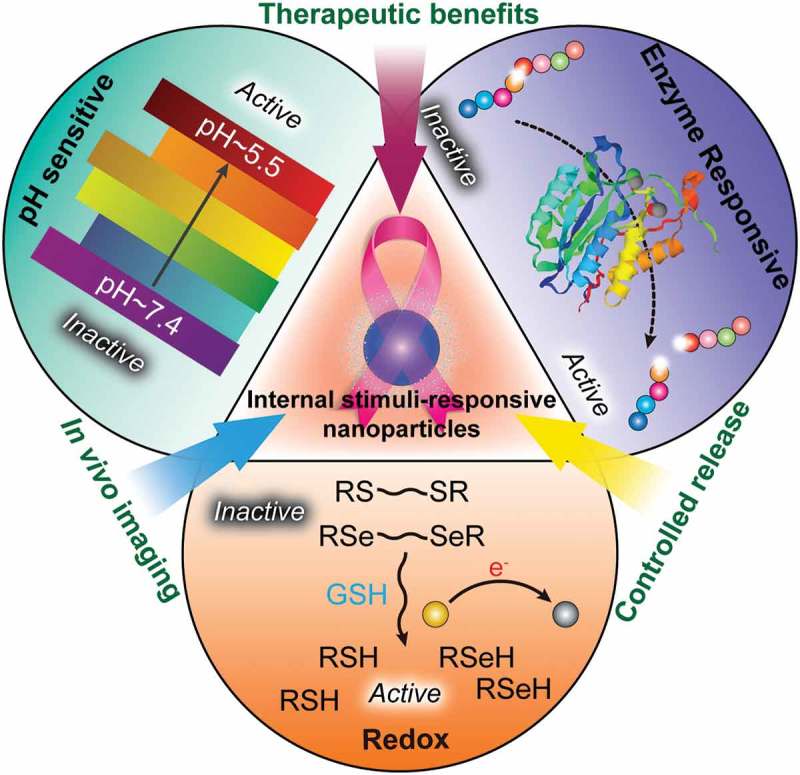
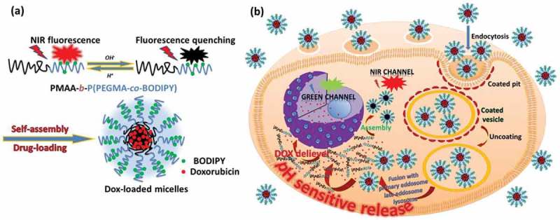
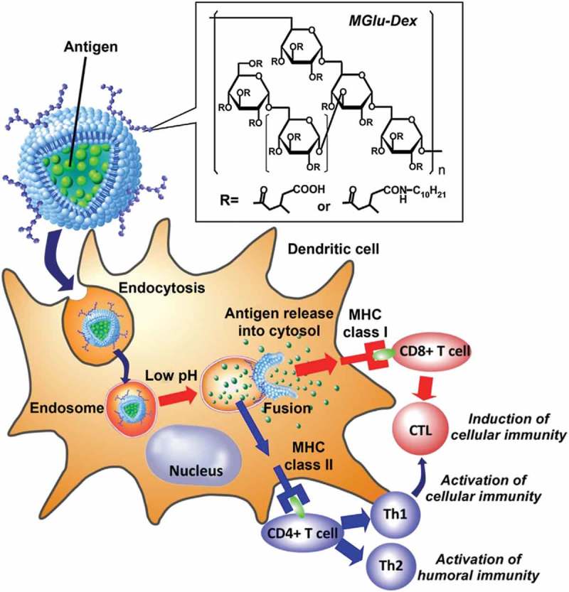
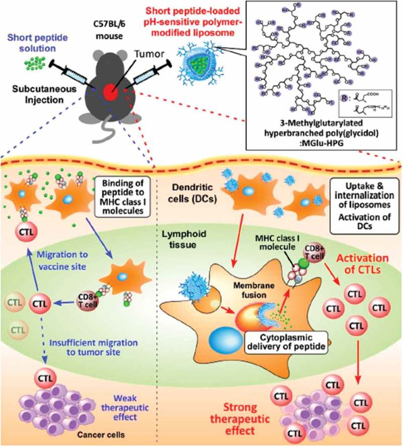
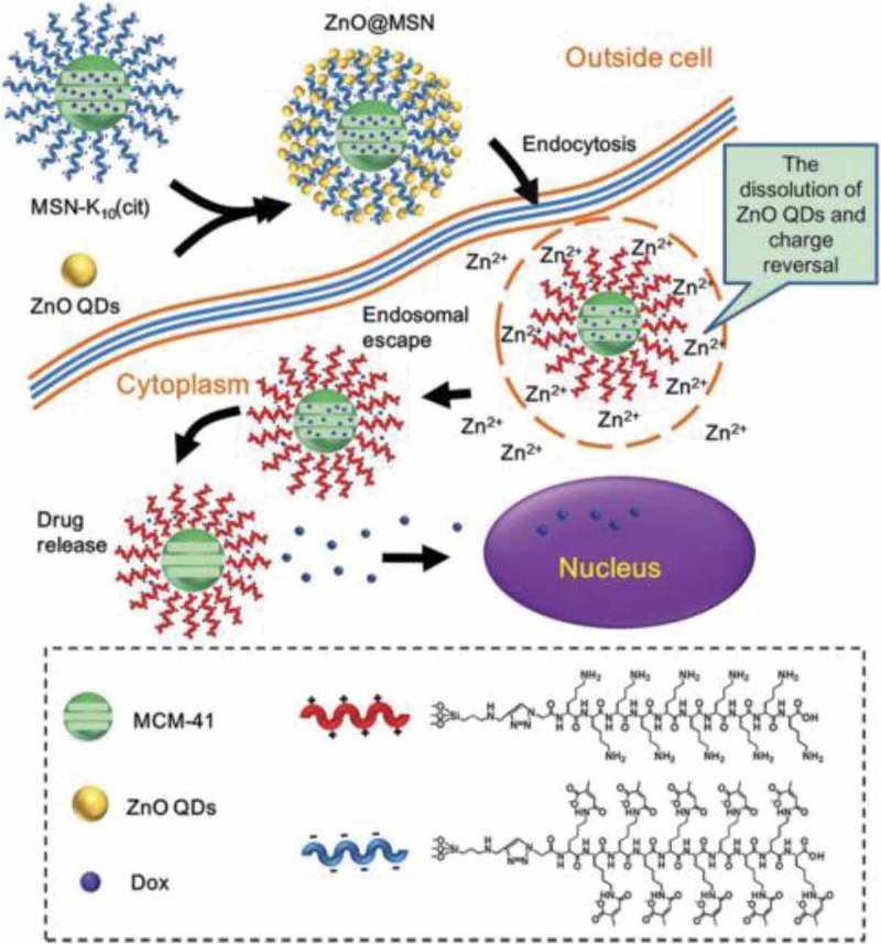
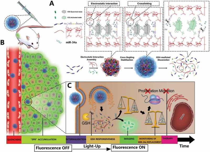
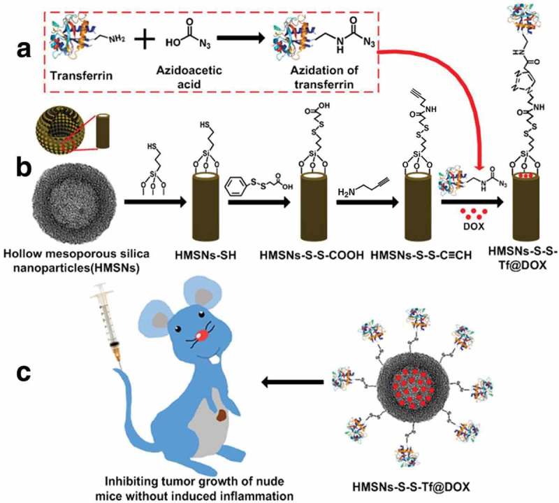
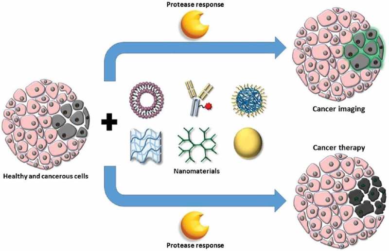
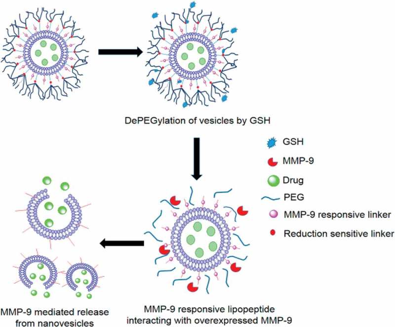
Nanotechnology has been extensively utilized in the design and development of powerful strategies for drug delivery and cancer theranostic. Nanoplatforms as a drug delivery system have many advantages such as in vivo imaging, combined drug delivery, extended circulation time, and systemic controlled release. The functional biomimetic drug delivery could be realized by incorporating stimuli-responsive (pH, temperature, redox potential, etc.) properties into the nanocarrier system, allowing them to bypass biological barriers and arrive at the targeted area. In this review, we discuss the role of internal stimuli-responsive nanocarrier system for imaging and drug delivery in cancer therapy. The development of internal stimuli-responsive nanoparticles is highlighted for precision drug delivery applications, with a particular focus on in vivo imaging, drug release performance, and therapeutic benefits.

Keywords: 10 Engineering and Structural materials; 211 Scaffold / Tissue engineering / Drug delivery; Functional nanoparticles; drug delivery; stimuli-responsive; theranostics.


Figures

None
Graphical abstract
Figure 1.
Figure 1.
Development of functional biomimetic nanoparticles for precision drug delivery applications in cancer treatment.
Figure 2.
Figure 2.
(a) Schematics of pH-modulation of fluorescence emission changes of PMAA-b-P(PEGMA-co-BODIPY) and its self-assembled multifunctional micelles. (b) Illustration of pH-sensitive drug-loaded micelles based on the PMAA-b-P(PEGMA-co-BODIPY) block copolymer for the intracellular release of DOX triggered by the acidic microenvironment. Reproduced with permission from Ref. [54]. Copyright 2015 Royal Society of Chemistry.
Figure 3.
Figure 3.
Design of MGlu-Dex-modified liposomes for induction of antigen-specific immunity. MGlu-Dex-modified liposome is taken up by DC via endocytosis and trapped in endosome. CTL stands for cytotoxic T lymphocyte. Its weakly acidic environment triggers destabilization of the liposome, which induces release of antigen molecules in endosome and their transfer to cytosol via fusion with endosome. Reproduced with permission from Ref. [71]. Copyright 2013 Elsevier Ltd.
Figure 4.
Figure 4.
Schematic illustration of antigen delivery routes for free peptide and peptide-loaded pH-sensitive polymer-modified liposomes. Reproduced with permission from Ref. [87]. Copyright 2016 Creative Commons.
Figure 5.
Figure 5.
Schematic illustration of intracellular drug release from ZnO@MSN. Reproduced with permission from Ref. [95]. Copyright 2015 American Chemical Society.
Figure 6.
Figure 6.
Schematic illustration of the design, construction, and application of light-up CMINs for monitoring of miR-34a replacement efficacy and accurate imaging-guided cancer therapy. ICG and miR-34a were simultaneously co-embedded in the nanocomplexes and subsequently stabilized by intermolecular cross-linking referred to as CMINs, which could better protect the embedded miR-34a and ICG and insure their release especially when dissociation of nanocomplexes happens. (A) Chemical structures of the components and preparation procedures of CMINs. (B) Schematic of the accumulation of CMINs in tumor sites via EPR effect (passive targeting). (C) Schematic of GSH-mediated dissociation of CMINs and the release of ICG and miR-34a upon intracellular uptake of nanocomplexes. Reproduced with permission from Ref. [109]. Copyright 2018 Wiley.
Figure 7.
Figure 7.
(a, b) Fabrication of redox-trigged HMSNs by using a disulfide bond as the intermediate linker. (c) Illustration of the intracellular redox-trigged HMSNs for targeted tumor therapy in vitro and in vivo. Reproduced with permission from Ref. [110]. Copyright Ivyspring International Publisher.
Figure 8.
Figure 8.
Tissues containing healthy (pink) and tumor (gray) cells can be treated with various nanomaterials, such as (from left to right) liposomes, protein-conjugates, polymeric nanoparticles, hydrogels, dendrimers, and inorganic metal nanoparticles, to deliver imaging agents or anticancer drugs with improved selectivity to tumor cells by incorporation of protease-responsiveness into the design of nanomaterials. Reproduced with permission from Ref. [128]. Copyright 2017 American Chemical Society.
Figure 9.
Figure 9.
Schematic representation of nanovesicles incorporating MMP-9 substrate lipopeptides and reduction-sensitive POPE-SS-PEG which render the nanovesicles responsive to extracellular, elevated levels of MMP-9 and GSH. Reproduced with permission from Ref. [130]. Copyright 2014 American Chemical Society.
Figure 10.
Figure 10.
In vivo biodistribution and in vitro fluorescence images of major organs and tumors. (A) In vivo time-dependent whole body fluorescence imaging of A549 tumor-bearing mice after intravenous injection of probes. (B) Quantification of average fluorescence signals in the tumor site of panel (A). (C) In vitro fluorescence images of major organs and tumors of mice after intravenous injection of probes over a period of 36 h. (D) Quantification of average fluorescence signals of panel C. Reproduced with permission from Ref. [131]. Copyright 2016 American Chemical Society.

References

    1. Neal JW, Sledge GW.. Decade in review-targeted therapy: successes, toxicities and challenges in solid tumours. Nat Rev Clin Oncol. 2014;11:627–628. - PubMed
    1. Metzger-Filho O, Moulin C, Awada A. Molecular targeted therapy in prevalent tumors: learning from the past and future perspectives. Curr Clin Pharmacol. 2010;5:166–177. - PubMed
    1. DeVita VT Jr., Chu E. A history of cancer chemotherapy. Cancer Res. 2008;68:8643–8653. - PubMed
    1. Cho K, Wang X, Nie S, et al. Therapeutic nanoparticles for drug delivery in cancer. Clin Cancer Res. 2008;14:1310–1316. - PubMed
    1. Parveen S, Sahoo SK. Polymeric nanoparticles for cancer therapy. J Drug Target. 2008;16:108–123. - PubMed

LinkOut - more resources