

Visual Explanations From Deep 3D Convolutional Neural Networks for Alzheimer's Disease Classification
- PMID: 30815203
- PMCID: PMC6371279
Visual Explanations From Deep 3D Convolutional Neural Networks for Alzheimer's Disease Classification
Abstract
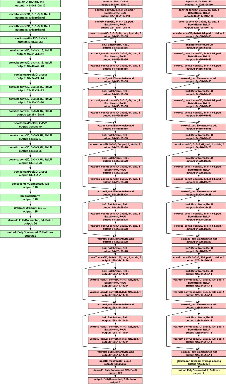
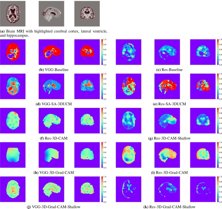
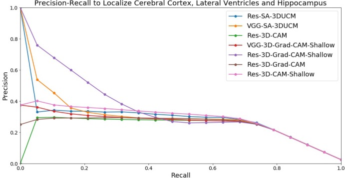
We develop three efficient approaches for generating visual explanations from 3D convolutional neural networks (3D-CNNs) for Alzheimer's disease classification. One approach conducts sensitivity analysis on hierarchical 3D image segmentation, and the other two visualize network activations on a spatial map. Visual checks and a quantitative localization benchmark indicate that all approaches identify important brain parts for Alzheimer's disease diagnosis. Comparative analysis show that the sensitivity analysis based approach has difficulty handling loosely distributed cerebral cortex, and approaches based on visualization of activations are constrained by the resolution of the convo-lutional layer. The complementarity of these methods improves the understanding of 3D-CNNs in Alzheimer's disease classification from different perspectives.
Figures
References
-
- Xu Jiaquan, Murphy Sherry L, Kochanek Kenneth D, Arias Elizabeth. Mortality in the united states. 2015. 2016. - PubMed
-
- McKhann Guy M, Knopman David S, Chertkow Howard, Hyman Bradley T, Jack Clifford R, Kawas Claudia H, Klunk William E, Koroshetz Walter J, Manly Jennifer J, Mayeux Richard, et al. The diagnosis of dementia due to alzheimers disease: Recommendations from the national institute on aging-alzheimers association workgroups on diagnostic guidelines for alzheimer’s disease. Alzheimer’s & dementia: the journal of the Alzheimer’s Association. 2011;7(3):263–269. - PMC - PubMed
-
- Khvostikov Alexander, Aderghal Karim, Benois-Pineau Jenny, Krylov Andrey, Catheline Gwenaelle. arXivpreprint arXiv:1801.05968. 2018. 3d cnn-based classification using smri and md-dti images for alzheimer disease studies.
-
- Korolev Sergey, Safiullin Amir, Belyaev Mikhail, Dodonova Yulia. Biomedical Imaging (ISBI2017), 2017 IEEE 14th International Symposium on. IEEE; 2017. Residual and plain convolutional neural networks for 3d brain mri classification.
Publication types
MeSH terms
LinkOut - more resources
Full Text Sources
Medical