

Extended resuscitative endovascular balloon occlusion of the aorta (REBOA)-induced type 2 myocardial ischemia: a time-dependent penalty
- PMID: 30815536
- PMCID: PMC6361364
- DOI: 10.1136/tsaco-2018-000194
Extended resuscitative endovascular balloon occlusion of the aorta (REBOA)-induced type 2 myocardial ischemia: a time-dependent penalty
Abstract
Background: Resuscitative Endovascular Balloon Occlusion of the Aorta (REBOA) increases cardiac-afterload and is used for patients in hemorrhagic shock. The cardiac tolerance of prolonged afterload augmentation in this context is unknown. The aim of this study is to quantify cardiac injury, if any, following 2, 3 and 4 hours of REBOA.
Methods: Anesthetized swine (70-90 kg) underwent a 40% controlled hemorrhage, followed by supraceliac resuscitative endovascular balloon occlusion of the aorta (REBOA) for 2 (n=5), 3 (n=5), and 4 hours (n=5). High-fidelity arterial wave form data were collected, and signal processing techniques were used to extract key inflection points. The adjusted augmentation index (AIx@75; augmentation pressure/pulse pressure, normalized for heart rate) was derived for use as a measure of aortic compliance (higher ratio = less compliance). Endpoints consisted of electrocardiographic, biochemical, and histologic markers of myocardial injury/ischemia. Regression modeling was used to assess the trend against time.
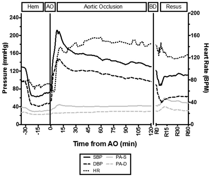
Results: All animals tolerated instrumentation, hemorrhage, and REBOA. The mean (±SD) systolic blood pressure (mm Hg) increased from 65±11 to 212±39 (p<0.001) during REBOA. The AIx@75 was significantly higher during REBOA than baseline, hemorrhage, and resuscitation phases (p<0.05). A time-dependent rise in troponin (R2=0.95; p<0.001) and T-wave deflection (R2=0.64; p<0.001) was observed. The maximum mean troponin (ng/mL) occurred at 4 hours (14.6±15.4) and maximum T-wave deflection (mm) at 65 minutes (3.0±1.8). All animals demonstrated histologic evidence of acute injury with increasing degrees of cellular myocardial injury.
Discussion: Prolonged REBOA may result in type 2 myocardial ischemia, which is time-dependent. This has important implications for patients where prolonged REBOA may be considered beneficial, and strategies to mitigate this effect require further investigation.
Level of evidence: II.
Keywords: aortic occlusion; arterial waveform; cardiac; cardiac injury; hemorrhage; ischemia; reboa; resuscitative endovascular balloon occlusion of the aorta.
Conflict of interest statement
Competing interests: JM is a clinical advisory board member for Prytime Medical.
Figures


References
-
- Wasicek PJ, Shanmuganathan K, Teeter WA, Gamble WB, Hu P, Stein DM, Scalea TM, Brenner ML. Assessment of blood flow patterns distal to aortic occlusion using CT in patients with resuscitative endovascular balloon occlusion of the aorta. J Am Coll Surg 2018;226:294–308. 10.1016/j.jamcollsurg.2017.12.005 - DOI - PubMed
-
- Manley JD, Mitchell BJ, DuBose JJ, Rasmussen TE. A modern case series of resuscitative endovascular balloon occlusion of the aorta (REBOA) in an out-of-hospital, combat casualty care setting. J Spec Oper Med 2017;17:1–8. - PubMed
-
- Sadek S, Lockey DJ, Lendrum RA, Perkins Z, Price J, Davies GE. Resuscitative endovascular balloon occlusion of the aorta (REBOA) in the pre-hospital setting: an additional resuscitation option for uncontrolled catastrophic haemorrhage. Resuscitation 2016;107:135–8. 10.1016/j.resuscitation.2016.06.029 - DOI - PubMed
LinkOut - more resources
Full Text Sources
Research Materials