Review
. 2019 May 6;14(9):889-906.
doi: 10.1002/cmdc.201900107. Epub 2019 Mar 26.

Covalent Inhibition in Drug Discovery

Affiliations
Review

Covalent Inhibition in Drug Discovery

Avick Kumar Ghosh et al. ChemMedChem. .

Abstract

Although covalent inhibitors have been used as therapeutics for more than a century, there has been general resistance in the pharmaceutical industry against their further development due to safety concerns. This inclination has recently been reverted after the development of a wide variety of covalent inhibitors to address human health conditions along with the US Food and Drug Administration (FDA) approval of several covalent therapeutics for use in humans. Along with this exciting resurrection of an old drug discovery concept, this review surveys enzymes that can be targeted by covalent inhibitors for the treatment of human diseases. We focus on protein kinases, RAS proteins, and a few other enzymes that have been studied extensively as targets for covalent inhibition, with the aim to address challenges in designing effective covalent drugs and to provide suggestions in the area that have yet to be explored.

Keywords: RAS proteins; acetylcholinesterase; caspase; cathepsin; covalent inhibitors; protein kinases.


Figures

Figure 1.
Figure 1.
Early covalent drugs. a) The action mechanism of aspirin; b) COX-1 active site serine and its covalent adduct after its reaction with c) a bromo derivative of aspirin; d) The action mechanism of penicillin; e) Co-crystal structure of DD-transpeptidase bound covalently with ampicillin (PDB entry: 5HL9) f) The amphicillin-serine complex in the active site of DD-transpeptidase. g) The action mechanism of acetaminophen.
Figure 2.
Figure 2.
A brief timeline of covalent drug discovery. Structures of covalent inhibitors are provided along with the enzymes/proteins they inhibit.
Figure 3.
Figure 3.
a) Two reversible EGFR inhibitors, Gefitinib and Erlotinib; b) First generation irreversible covalent inhibitors of EGFR, PD168393, PF00299804 and EKB569. All contain an acrylamide moiety, highlighted in a box, as an electrophilic warhead. c) Neratinib and its complex with EGFR T790M (PDB entry: 2JIV). Gatekeeper residue (Met790) is shown in red. d) Afatinib and its complex with EGFR T790M (PDB entry: 4G5P).
Figure 4.
Figure 4.
Third generation irreversible covalent EGFR inhibitors. a) WZ4002 and it complex with EGFR T790M (PDB entry: 3IKA); b) Osimertinib and its complex with wild type EGFR (PDB entry: 4ZAU); c) PF-06459988 and its complex with EGFR L858R/T790M (PDB entry: 5GH7); d) Other third generation irreversible covalent EGFR inhibitors.
Figure 5.
Figure 5.
Covalent BTK inhibitors. a) Ibrutinib; b) A pyrrolopyrimidine precursor of ibrutinib and its complex with BTK (PDB entry: 3GEN); c) Acalabrutinib; d) Second generation irreversible covalent BTK inhibitors.
Figure 6.
Figure 6.
a) PP1, a Src family kinase inhibitor; b) FMK, an irreversible covalent inhibitor of RSK2; c) Co-crystal structure of the SRc family kinase HCK bound to PP1 (PDB entry: 1QCF) with N2 properly oriented to Val 281 (shown in green) which corresponds to C436 in RSK2.
Figure 7.
Figure 7.
a) The FIIN series inhibitors of FGFR and the co-crystal structure of FIIN3 complexed with FGFR V550L (PDB entry: 4R6V); b) PRN1371, a covalent FGFR inhibitor; c) Non-covalent CDK2 inhibitor NU6102, its corresponding covalent CDK2 inhibitor NU6300, and a co-crystal structure of CDK2 bound covalently with NU6300 (PDB entry: 5CYI) via K89; d) Covalent CDK7 inhibitor THZ1, THZ351, non-covalent CDK12 and CDK13 inhibitor THZ531, and a co-crystal structure of CDK12 bound covalently with THZ531 (PDB entry: 5ACB); e) PI3Kα inhibitor CNX-1351.
Figure 8.
Figure 8.
a) Reversible covalent inhibitors CN-NHiPr and CN-NHtBu of RSK2 that were derived from FMN and a co-crystal structure of RSK2 bound covalently with CN-NHtBu at C436 (PDB entry: 4D9U); b) Cyanoarylamide-based reversible covalent inhibitors of BTK and a co-crystal structure of BTK bound covalently with compound 3 at C481.
Figure 9.
Figure 9.
Tethering and electrophilic compounds that selectively bind to oncogenic KRAS G12C. a) Tethering compounds that binds covalently to KRAS G12C; b) Crystal structure of the KRAS G12C complex with compound 6 (PDB entry: 4LUC); c) Electrophiles with the vinyl sulphonamide moiety that bind covalently to KRAS G12C; d) Crystal structure of the KRAS G12C complex with compound 12; e) Electrophiles with the acrylamide moiety that bind covalently to KRAS G12C (PDB entry: 4LYF).
Figure 10.
Figure 10.
Some other covalent inhibitors of KRAS G12C. a. SML-8–73-1; b. The structure of KRAS G12C bound with SML-8–73-1 (PDB entry: 4NMM); c. SML-10–70-1; d. ARS-853; e. The structure of KRAS G12C bound with ARS-853 (PDB entry: 5F2E).
Figure 11.
Figure 11.
a. AchE inhibitors; b. Structure of mouse AChE; c. AChE with choline bound at the active site (PDB entry: 2HA3); d. Interactions of rivastigmine with AChE in the active site.
Figure 12.
Figure 12.
Covalent cathepsin inhibitors. a. Balicatib; b. Odonacatib; c. The covalent adduct between odonacatib and Cys25 in the CatK active site (PDB entry: 5TDI); d. JPM-OEt; e. JPM-565.
Figure 13.
Figure 13.
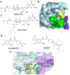
Covalent caspase inhibitors. a-b. A covalent caspase 8 inhibitor and its caspase 8 adduct structure (PDB entry: 3KJN); c. Three covalent caspase 3 and caspase 6 inhibitors that contain the tetrafluorophenyl ether moeity; d. Caspase 3 inhibitors that are 2-acetic acid derivatives.
Figure 14.
Figure 14.
a. Thiol containing allosteric inhibitors of caspase enzymes; b. Crystal structure of caspase 7 bound with compound 1 (PDB entry:1SHL); c. an enlarged view at the active site for the structure shown in b.
Figure 15.
Figure 15.
Structures of orlistat, beloranib, vildagliptin, and Saxagliptin.

References

    1. Baillie TA, Angew Chem Int Ed Engl 2016, 55, 13408–13421. - PubMed
    1. Singh J, Petter RC, Baillie TA, Whitty A, Nat Rev Drug Discov 2011, 10, 307–317. - PubMed
    1. Roth GJ, Stanford N, Majerus PW, Proc Natl Acad Sci U S A 1975, 72, 3073–3076. - PMC - PubMed
    1. Picot D, Loll PJ, Garavito RM, Nature 1994, 367, 243–249; P. J. Loll, D. Picot, R. M. Garavito, Nat Struct Biol 1995, 2, 637–643. - PubMed
    1. Baillie TA, Drug Metab Rev 2015, 47, 4–11. - PubMed

Publication types

LinkOut - more resources