. 2019 Feb 28;9(1):2996.
doi: 10.1038/s41598-018-35840-3.

Migrated T lymphocytes into malignant pleural effusions: an indicator of good prognosis in lung adenocarcinoma patients

Affiliations

Migrated T lymphocytes into malignant pleural effusions: an indicator of good prognosis in lung adenocarcinoma patients

Juan C Nieto et al. Sci Rep. .

Abstract

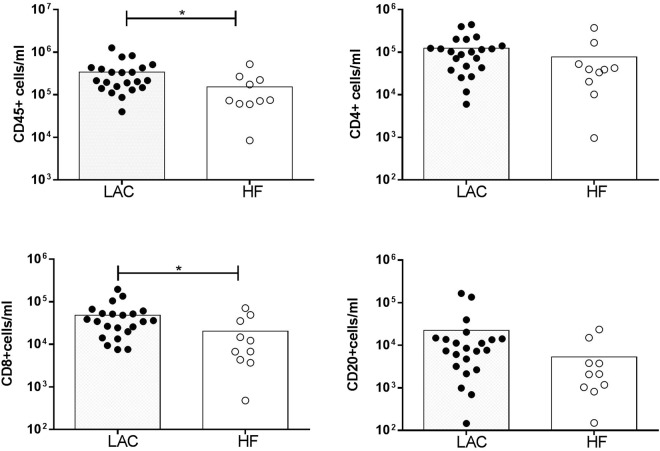
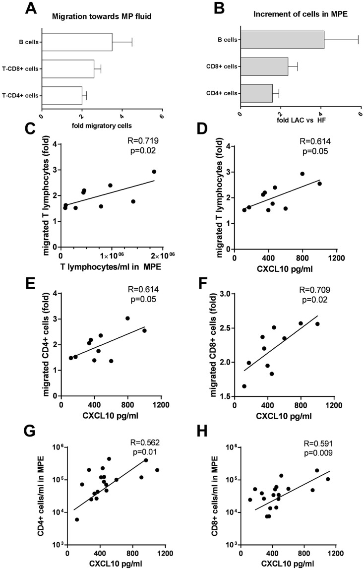
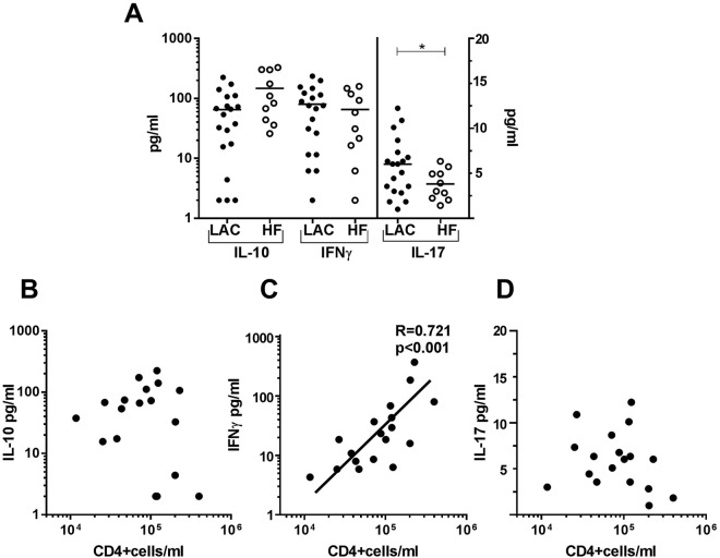
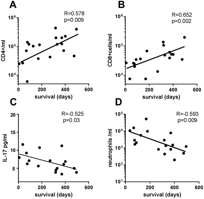
The presence of leukocyte subpopulations in malignant pleural effusions (MPEs) can have a different impact on tumor cell proliferation and vascular leakiness, their analysis can help to understand the metastatic microenvironment. We analyzed the relationship between the leukocyte subpopulation counts per ml of pleural fluid and the tumor cell count, molecular phenotype of lung adenocarcinoma (LAC), time from cancer diagnosis and previous oncologic therapy. We also evaluated the leukocyte composition of MPEs as a biomarker of prognosis. We determined CD4+ T, CD8+ T and CD20+ B cells, monocytes and neutrophils per ml in pleural effusions of 22 LAC and 10 heart failure (HF) patients by flow cytometry. Tumor cells were identified by morphology and CD326 expression. IFNγ, IL-10 and IL-17, and chemokines were determined by ELISAs and migratory response to pleural fluids by transwell assays. MPEs from LAC patients had more CD8+ T lymphocytes and a tendency to more CD4+ T and CD20+ B lymphocytes than HF-related fluids. However, no correlation was found between lymphocytes and tumor cells. In those MPEs which were detected >1 month from LAC diagnosis, there was a negative correlation between pleural tumor cells and CD8+ T lymphocytes. CXCL10 was responsible for the attraction of CD20+ B, CD4+ T and CD8+ T lymphocytes in malignant fluids. Concentrations of IL-17 were higher in MPEs than in HF-related effusions. Survival after MPE diagnosis correlated positively with CD4+ T and CD8+ T lymphocytes, but negatively with neutrophils and IL-17 levels. In conclusion, lymphocyte enrichment in MPEs from LAC patients is mostly due to local migration and increases patient survival.


Conflict of interest statement

The authors declare no competing interests.

Figures

Figure 1
Figure 1
Leukocytes and lymphocyte subpopulations in malignant and non-malignant pleural effusions. Cells from MPEs of lung adenocarcinoma (LAC) and heart failure (HF) were stained with anti-CD3, CD4, CD14, CD16, CD20 and CD45 monoclonal antibodies to be analyzed by flow cytometry. Data are presented as mean. CD4+ cells/ml and CD20+ cells/ml in LAC and HF tended to be different (p = 0.04 and p = 0.06 respectively). Statistical analysis was performed using a non-parametric Mann-Whitney test. p < 0.05 was considered statistically significant. *p < 0.05.
Figure 2
Figure 2
Correlation between lymphocyte populations and tumor cells in malignant pleural effusions from lung cancer. Cells from MPEs of LAC patients were stained with anti-CD3, CD4, CD20, CD45 and EpCAM (CD326) monoclonal antibodies to be analyzed by flow cytometry. Tumor cells were identified as CD45- CD326 (EpCAM)+ cells. Correlations between CD4+ T and CD8+ T, CD4+ T and CD20+ T, and CD8+ T and CD20+ B lymphocytes cells/ml and between tumor cells/ml and CD4+ T, CD8+ T and CD20+ B lymphocytes cells/ml were analyzed by Spearman’s correlation. Black circles correspond to LAC patients diagnosed with cancer >1 month before of MPE formation. Statistically significant correlations (p < 0.05) are shown.
Figure 3
Figure 3
Association between migration of lymphocytes toward malignant pleural fluids and number of lymphocytes in malignant pleural effusions. Lysed whole blood cells from healthy donors (3 × 105 cells) were added to 3 μm insert transwells. Under insert, culture medium with 10% of FCS or medium with 10% of MP fluid was added to the well. After 4 h, cells were collected from the well and stained with anti-CD3 and CD4. (A) Migration folds of CD20+ B, CD8+ T, and CD4+ T lymphocytes in a medium with 10% MP fluid vs medium with 10% FCS are shown. (B) Increments of CD20+ B, CD8+ T and CD4+ T lymphocytes in MPE from LAC vs HF are shown. Correlation of fold of migrated T lymphocytes with (C) T lymphocytes cells per ml in MPE and with (D) CXCL10 levels (pg/ml) in MP fluids. Correlation of CXCL10 levels with the fold of (E) migrated CD4+ T and (F) CD8+ T lymphocytes and (G) CD4+ T lymphocytes and (H) CD8+ T lymphocytes per ml in MPE. We used Spearman’s correlation. Statistically significant correlations (p < 0.05) are shown.
Figure 4
Figure 4
IL-10, IFNγ and IL-17 levels in MPE from LAC and pleural fluids from HF and the relationship of cytokines with T CD4+ lymphocytes per ml in MPE. Pleural fluids LAC and HF patients were filtered with 0.22 μm. (A) OD levels of IL-10, IFNγ, and IL-17 determined by ELISA. IL-10 levels in LAC and HF tended to be different (p = 0.06). Mann-Whitney test was used for comparison between LAC and HF. Correlation of CD4+ cells/ml with OD of (B) IL-10, (C) IFNγ and (D) IL-17 was analyzed by Spearman’s correlation. p < 0.05 was considered statistically significant. *p < 0.05.
Figure 5
Figure 5
Association of cytokines and leukocytes per ml in MPE with survival days from MPE formation. Correlation between survival days after MPE formation and cells per ml of (A) CD4+ T, (B) CD8+ T, (C) IL-17 and (D) neutrophils per ml was analyzed by Spearman’s correlation. p < 0.05 was considered statistically significant.

References

    1. Porcel JM, Light RW. Pleural effusions. Dis. Mon. 2013;59:29–57. doi: 10.1016/j.disamonth.2012.11.002. - DOI - PubMed
    1. Porcel JM, Esquerda A, Vives M, Bielsa S. Etiology of pleural effusions: analysis of more than 3,000 consecutive thoracenteses. Arch. Bronconeumol. 2014;50:161–165. doi: 10.1016/j.arbres.2013.11.007. - DOI - PubMed
    1. Porcel JM, et al. Clinical features and survival of lung cancer patients with pleural effusions. Respirology. 2015;20:654–659. doi: 10.1111/resp.12496. - DOI - PubMed
    1. Marazioti A, Blackwell TS, Stathopoulos GT. The lymphatic system in malignant pleural effusion. Drain or immune switch? Am. J. Respir. Crit. Care Med. 2014;189:626–627. doi: 10.1164/rccm.201401-0140ED. - DOI - PubMed
    1. Psallidas I, et al. Secreted phosphoprotein-1 directly provokes vascular leakage to foster malignant pleural effusion. Oncogene. 2013;32:528–535. doi: 10.1038/onc.2012.57. - DOI - PubMed

Publication types

MeSH terms