

Distribution and Morphology of Cortical Terminals in the Cat Thalamus from the Anterior Ectosylvian Sulcus
- PMID: 30816175
- PMCID: PMC6395774
- DOI: 10.1038/s41598-019-39327-7
Distribution and Morphology of Cortical Terminals in the Cat Thalamus from the Anterior Ectosylvian Sulcus
Abstract
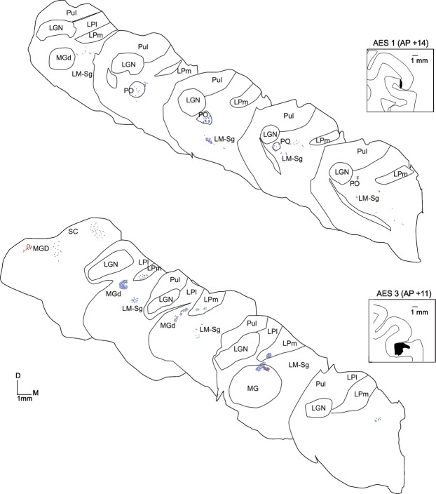
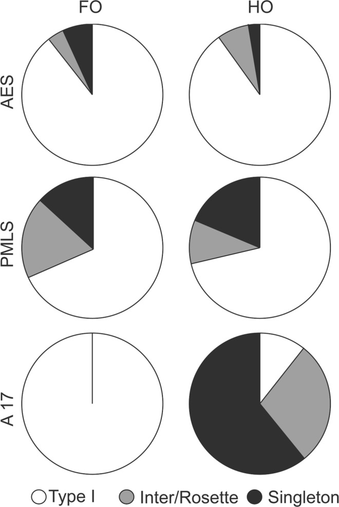
Two main types of cortical terminals have been identified in the cat thalamus. Large (type II) have been proposed to drive the response properties of thalamic cells while smaller (type I) are believed to modulate those properties. Among the cat's visual cortical areas, the anterior ectosylvian visual area (AEV) is considered as one of the highest areas in the hierarchical organization of the visual system. Whereas the connections from the AEV to the thalamus have been recognized, their nature (type I or II) is presently not known. In this study, we assessed and compared the relative contribution of type I and type II inputs to thalamic nuclei originating from the AEV. The anterograde tracer BDA was injected in the AEV of five animals. Results show that (1) both type I and II terminals from AEV are present in the Lateral Posterior- Pulvinar complex, the lateral median suprageniculate complex and the medial and dorsal geniculate nuclei (2) type I terminals significantly outnumber the type II terminals in almost all nuclei studied. Our results indicate that neurons in the AEV are more likely to modulate response properties in the thalamus rather than to determine basic organization of receptive fields of thalamic cells.
Conflict of interest statement
The authors declare no competing interests.
Figures


Similar articles
-
Distribution, morphology, and synaptic targets of corticothalamic terminals in the cat lateral posterior-pulvinar complex that originate from the posteromedial lateral suprasylvian cortex.J Comp Neurol. 2006 Aug 20;497(6):847-63. doi: 10.1002/cne.21024. J Comp Neurol. 2006. PMID: 16802329 Free PMC article.
-
Connections of the anterior ectosylvian visual area (AEV).Exp Brain Res. 1986;62(2):225-40. doi: 10.1007/BF00238842. Exp Brain Res. 1986. PMID: 3709710
-
Physiologic and anatomic investigation of a visual cortical area situated in the ventral bank of the anterior ectosylvian sulcus of the cat.Exp Brain Res. 1982;46(1):1-11. doi: 10.1007/BF00238092. Exp Brain Res. 1982. PMID: 7067781
-
Insular cortex and neighboring fields in the cat: a redefinition based on cortical microarchitecture and connections with the thalamus.J Comp Neurol. 1997 Aug 4;384(3):456-82. doi: 10.1002/(sici)1096-9861(19970804)384:3<456::aid-cne10>3.0.co;2-h. J Comp Neurol. 1997. PMID: 9254039
-
The dorsal lateral geniculate nucleus and the pulvinar as essential partners for visual cortical functions.Front Neurosci. 2023 Aug 30;17:1258393. doi: 10.3389/fnins.2023.1258393. eCollection 2023. Front Neurosci. 2023. PMID: 37712093 Free PMC article. Review.
Cited by
-
Hierarchical Organization of Corticothalamic Projections to the Pulvinar.Cereb Cortex Commun. 2020 Jul 7;1(1):tgaa030. doi: 10.1093/texcom/tgaa030. eCollection 2020. Cereb Cortex Commun. 2020. PMID: 34296104 Free PMC article.
-
Corticothalamic Projections Gate Alpha Rhythms in the Pulvinar.Front Cell Neurosci. 2021 Dec 6;15:787170. doi: 10.3389/fncel.2021.787170. eCollection 2021. Front Cell Neurosci. 2021. PMID: 34938163 Free PMC article.
References
Publication types
MeSH terms
Grants and funding
LinkOut - more resources
Full Text Sources
Miscellaneous
