

Analysis of the Antigenic Properties of Membrane Proteins of Mycobacterium tuberculosis
- PMID: 30816178
- PMCID: PMC6395656
- DOI: 10.1038/s41598-019-39402-z
Analysis of the Antigenic Properties of Membrane Proteins of Mycobacterium tuberculosis
Abstract
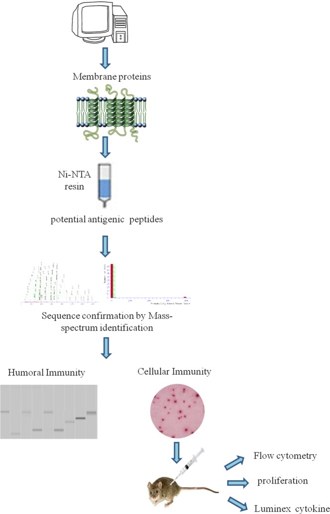
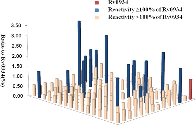
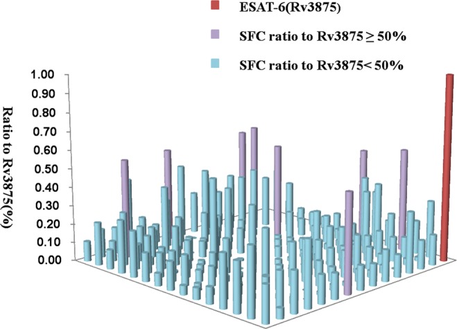
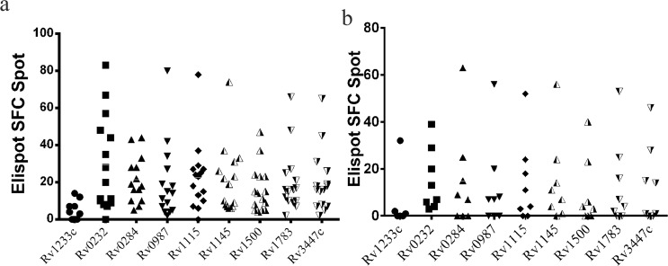
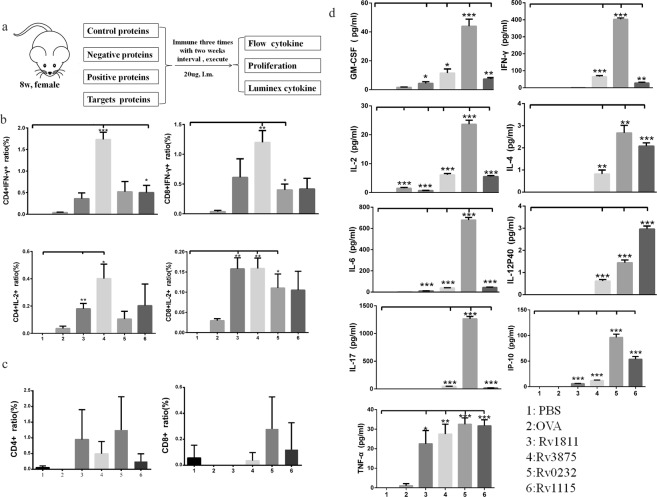
Tuberculosis (TB) is a continuing major threat to global health and a leading cause of death, particularly in developing countries. In this study, we aimed to identify a specific and sensitive diagnostic biomarker and develop a vaccine to prevent this disease. We investigated membrane proteins to reveal biomarkers in serum and peripheral blood mononuclear cells (PBMCs) obtained from TB patients. We employed Western blotting to evaluate serological immunoglobulin G levels, and Enzyme Linked Immunospot (ELISpot) to assess the antigen-specific cellular interferon-γ secretion from PBMCs after membrane protein stimulation. A total of 219 membrane proteins were identified, 52 exhibited at a higher levels than the 38-kDa prositive control. Of these 18 exhibited reacted ratios above 1, especially Rv1111c (427-981), with a ratios at 3.38. Accuracy and sensitivity were markedly higher for the top two antigen candidates, Rv0232 and Rv1115, after two rounds of ELISpot tests than ESAT-6 in the commercial kit (42.15 and 43.62%, respectively). These two proteins were administered to mice to detect whether they acted as effective antigens in vivo. These data provide a comprehensive view of the membranes involved in humoural and cellular immune responses that may be used as biomarkers for TB and candidates for a vaccine.
Conflict of interest statement
The authors declare no competing interests.
Figures
Similar articles
-
Exploration of novel cellular and serological antigen biomarkers in the ORFeome of Mycobacterium tuberculosis.Mol Cell Proteomics. 2014 Mar;13(3):897-906. doi: 10.1074/mcp.M113.032623. Epub 2014 Jan 21. Mol Cell Proteomics. 2014. PMID: 24447912 Free PMC article.
-
Identification and evaluation of the novel immunodominant antigen Rv2351c from Mycobacterium tuberculosis.Emerg Microbes Infect. 2017 Jun 7;6(6):e48. doi: 10.1038/emi.2017.34. Emerg Microbes Infect. 2017. PMID: 28588287 Free PMC article.
-
The diagnostic potential of MPT63-derived HLA-A*0201-restricted CD8+ T-cell epitopes for active pulmonary tuberculosis.Microbiol Immunol. 2015 Dec;59(12):705-15. doi: 10.1111/1348-0421.12339. Microbiol Immunol. 2015. PMID: 26577013
-
Vaccines for Leprosy and Tuberculosis: Opportunities for Shared Research, Development, and Application.Front Immunol. 2018 Feb 26;9:308. doi: 10.3389/fimmu.2018.00308. eCollection 2018. Front Immunol. 2018. PMID: 29535713 Free PMC article. Review.
-
Genome wide approaches discover novel Mycobacterium tuberculosis antigens as correlates of infection, disease, immunity and targets for vaccination.Semin Immunol. 2018 Oct;39:88-101. doi: 10.1016/j.smim.2018.07.001. Epub 2018 Jul 7. Semin Immunol. 2018. PMID: 30327124 Review.
Cited by
-
Proteome Profile Changes Induced by Heterologous Overexpression of Mycobacterium tuberculosis-Derived Antigens PstS-1 (Rv0934) and Ag85B (Rv1886c) in Mycobacterium microti.Biomolecules. 2022 Dec 8;12(12):1836. doi: 10.3390/biom12121836. Biomolecules. 2022. PMID: 36551264 Free PMC article.
-
Patterns of T and B cell responses to Mycobacterium tuberculosis membrane-associated antigens and their relationship with disease activity in rheumatoid arthritis patients with latent tuberculosis infection.PLoS One. 2021 Aug 2;16(8):e0255639. doi: 10.1371/journal.pone.0255639. eCollection 2021. PLoS One. 2021. PMID: 34339423 Free PMC article.
-
Evaluation of the Toxic Activity of the Graphene Oxide in the Ex Vivo Model of Human PBMC Infection with Mycobacterium tuberculosis.Microorganisms. 2023 Feb 22;11(3):554. doi: 10.3390/microorganisms11030554. Microorganisms. 2023. PMID: 36985128 Free PMC article.
-
In-silico development of a novel TLR2-mediating multi-epitope vaccine against Mycobacterium tuberculosis.In Silico Pharmacol. 2025 Feb 25;13(1):34. doi: 10.1007/s40203-025-00322-8. eCollection 2025. In Silico Pharmacol. 2025. PMID: 40018380
-
From immunology to artificial intelligence: revolutionizing latent tuberculosis infection diagnosis with machine learning.Mil Med Res. 2023 Nov 28;10(1):58. doi: 10.1186/s40779-023-00490-8. Mil Med Res. 2023. PMID: 38017571 Free PMC article. Review.
References
-
- WHO. World malaria report 2011 (2011).
Publication types
MeSH terms
Substances
LinkOut - more resources
Full Text Sources
