. 2019 Feb 28;9(1):3161.
doi: 10.1038/s41598-019-39341-9.

Tissue Engineered Neural Constructs Composed of Neural Precursor Cells, Recombinant Spidroin and PRP for Neural Tissue Regeneration

Affiliations

Tissue Engineered Neural Constructs Composed of Neural Precursor Cells, Recombinant Spidroin and PRP for Neural Tissue Regeneration

V P Baklaushev et al. Sci Rep. .

Abstract

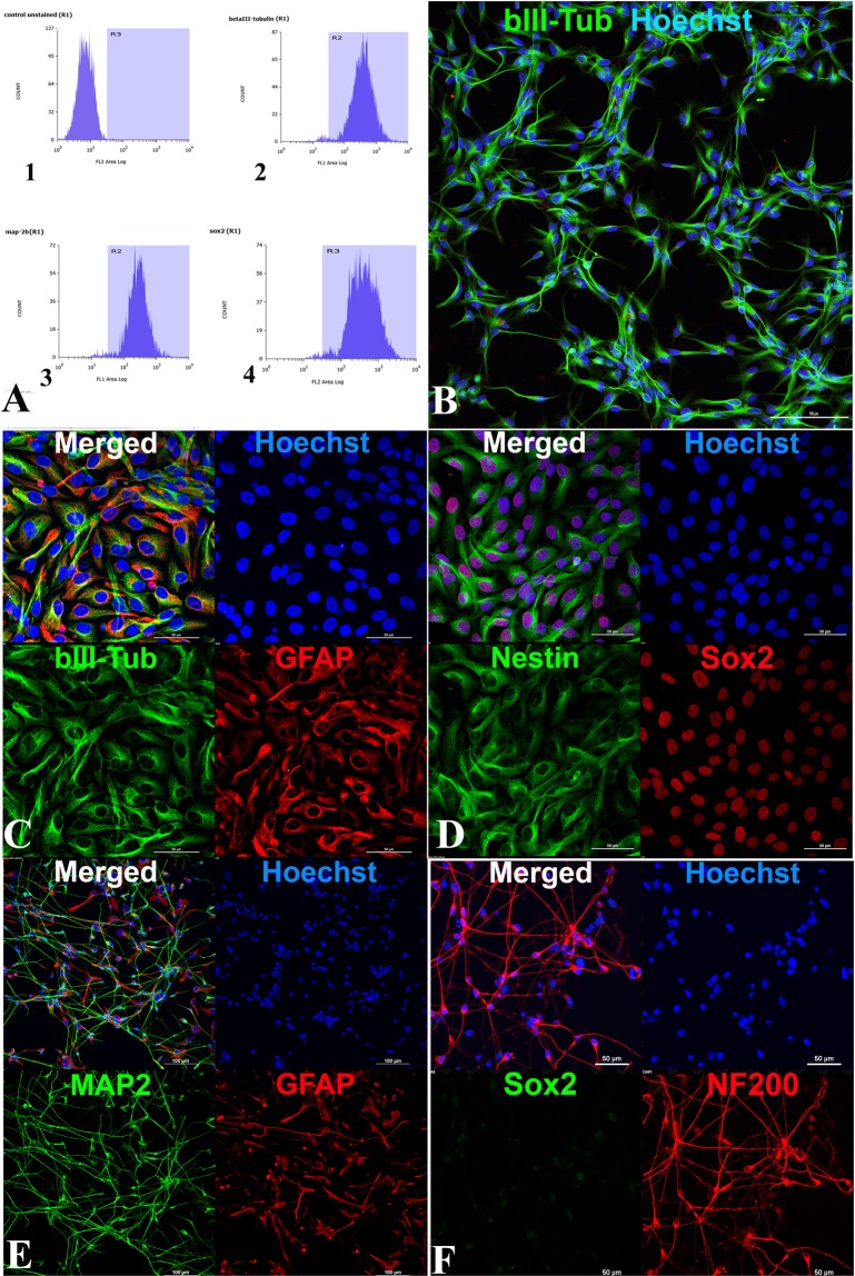
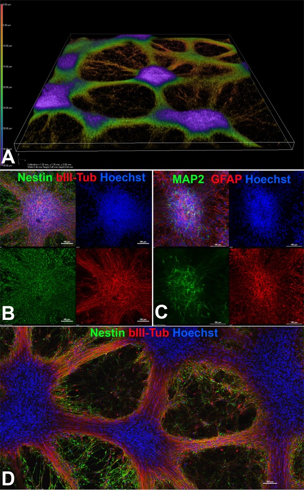
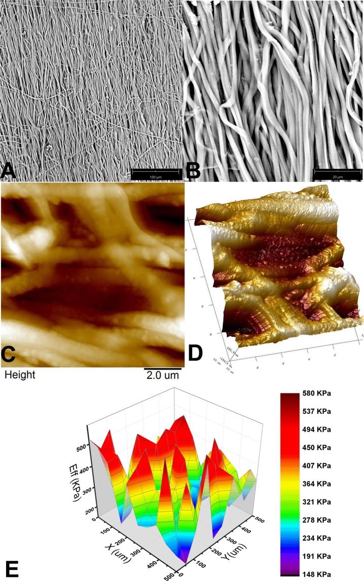
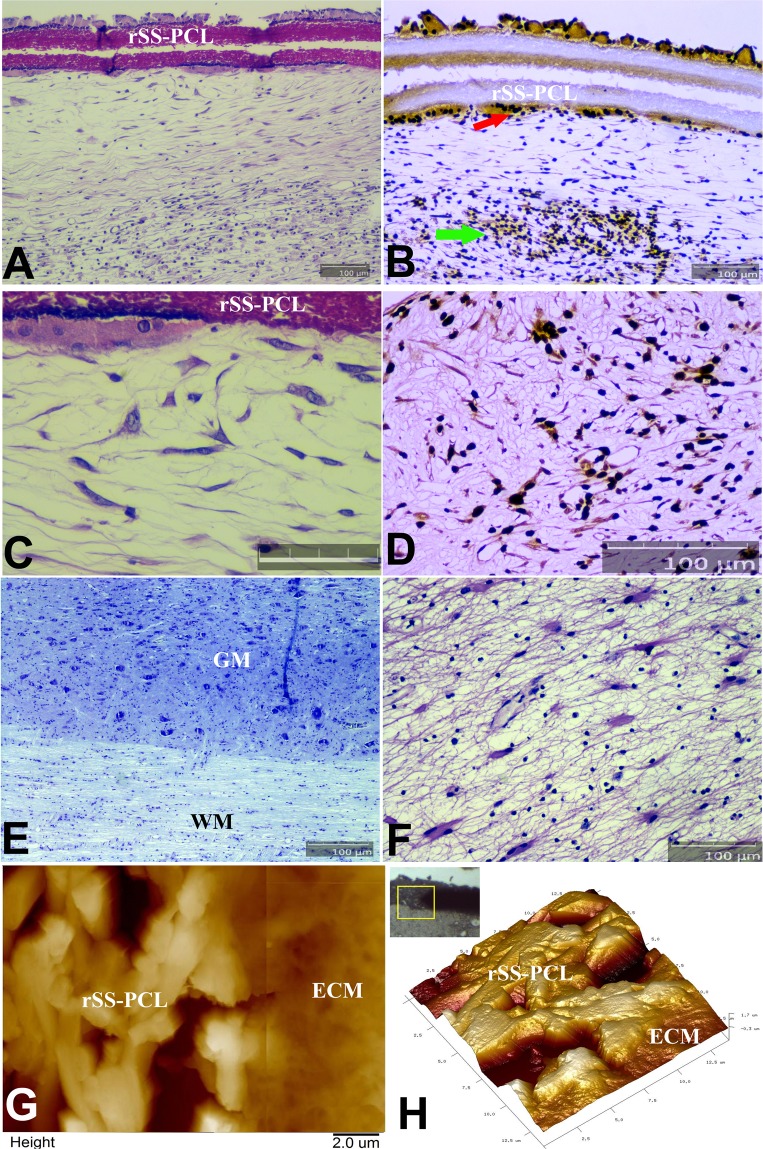
We have designed a novel two-component matrix (SPRPix) for the encapsulation of directly reprogrammed human neural precursor cells (drNPC). The matrix is comprised of 1) a solid anisotropic complex scaffold prepared by electrospinning a mixture of recombinant analogues of the spider dragline silk proteins - spidroin 1 (rS1/9) and spidroin 2 (rS2/12) - and polycaprolactone (PCL) (rSS-PCL), and 2) a "liquid matrix" based on platelet-rich plasma (PRP). The combination of PRP and spidroin promoted drNPC proliferation with the formation of neural tissue organoids and dramatically activated neurogenesis. Differentiation of drNPCs generated large numbers of βIII-tubulin and MAP2 positive neurons as well as some GFAP-positive astrocytes, which likely had a neuronal supporting function. Interestingly the SPRPix microfibrils appeared to provide strong guidance cues as the differentiating neurons oriented their processes parallel to them. Implantation of the SPRPix matrix containing human drNPC into the brain and spinal cord of two healthy Rhesus macaque monkeys showed good biocompatibility: no astroglial and microglial reaction was present around the implanted construct. Importantly, the human drNPCs survived for the 3 month study period and differentiated into MAP2 positive neurons. Tissue engineered constructs based on SPRPix exhibits important attributes that warrant further examination in spinal cord injury treatment.


Conflict of interest statement

The authors declare no competing interests.

Figures

Figure 1
Figure 1
Immunophenotyping of drNPC. (A) Cytofluorometry of drNPC marker expression. 1. Negative control (isotypic immunoglobulins) 2. βIII-tubulin 3. MAP2 and 4. SOX2. (BF) Immunocytochemical analysis for different markers (B) anti-βIII-tubulin. Bar = 100 µm (C,D) Undifferentiated drNPC coexpress βIII-tubulin (green) and GFAP (red) (C), as well as Nestin (green) and SOX2 (red) (D). (E,F) Differentiation of drNPC: MAP2-positive neurons (green) and GFAP-positive astrocytes (red) (E) NF200-positve (red) and SOX2 negative neurons (F). In panels (B–F), the blue channel corresponds to the Hoechst stained cell nuclei. In panels С–F Bar = 50 µm. Laser scanning confocal microscopy using Nikon A1 device.
Figure 2
Figure 2
drNPC cultured in the PRP based liquid matrix. (A) 3D reconstruction of cultured drNPCs-01 in PRP based hydrogel using laser scanning confocal microscopy. (B) Spontaneous differentiation of neural organoids formed in PRP based hydrogel with the appearance of βIII-tubulin neuronal progenitors (red) amongst the more common Nestin positive (green) NPCs (C) Neural organoids stained with MAP2 (green) and GFAP (red) further confirming glial and neuronal differentiation. (D) Low magnification image showing the overall morphology of the differentiating cultures with large numbers of Nestin positive (green) NPCs and βIII-tubulin positive neurons (red). The optical plane cuts through the center of the neural organoids. (B–D) bar = 100 µm.
Figure 3
Figure 3
The spatial architecture and mechanical properties of the rSS-PCL scaffold: (A,B) SEM images, (C), 2D and (D), 3D – atomic force microscopy images, (E). typical Young’s modulus distribution obtained by nanoindentation.
Figure 4
Figure 4
drNPC cultured in the rSS-PCL scaffold with or without the PRP-based liquid matrix (SPRPix matrix). (A–C) SPRPix (PRP + rSS-PCL scaffold); (A) Nestin (green), auto-fluorescence of the SPRPix matrix (red). (B) Spontaneous neuronal and glial differentiation revealed by MAP2 (green) and GFAP (red). (C) Intense proliferation of SOX2-positve (green) drNPCs, and βIII-tubulin-positive (red) neuronal progenitors. (D) The same concentration of drNPC cells seeded on rSS-PCL scaffold only. Nestin (green), βIII-tubulin (red). (E,F) high magnification examples of drNPCs cultured on rSS-PCL alone (E) or on SPRPix (F). Bar = 50 μm.
Figure 5
Figure 5
Analysis of structure alignment using image FFT and second order moments. (A) SEM of rSS-PCL scaffold; (B). MAP2 staining; drNPC in SPRPix matrix (rSS-PCL + PRP); (C). βIII-tubulin staining; drNPC in SPRPix matrix; (D). βIII-tubulin staining; drNPC in rSS-PCL scaffold; (E). βIII-tubulin staining; drNPC cultured on isotropic surface. Left – original grayscale images; Middle – FFT images with equivalent covariance ellipses; Right – angular distribution of FFT intensity with normal fit.
Figure 6
Figure 6
Histological and immunohistochemical (IHC) analysis of the endogenous immune response to the drNPC-SPRPix implanted into the brain cortex of a rhesus macaque. (A) Nissl stain; magnification ×100 (B,С). H&E stain of the interface between the SPRPix and the surrounding host tissue; magnifications ×100 (B) and ×200 (С). (D,E). Representative images of CD3 staining showing low level of T cell infiltration in the brain tissue adjacent to the drNPC-SPRPix implant. (FH) Representative images of CD68 staining showing examples of microglial/macrophages in the brain adjacent to the drNPC-SPRPix implant (G) and among the human cell graft (H). (E,H) depict the areas with the highest immune cell infiltration noted. Bar size 100 µm. Notations: rSS-PCL-spidroin-polycaprolactone scaffold; Ctx – brain cortex.
Figure 7
Figure 7
Immunofluorescence analysis of the brain of a rhesus macaque at the site of implantation of the SPRPix matrix with human drNPC. (A) Representative low magnification image of the site of drNPC-SPRPix implantantation showing staining by human specific anti-mitochondria antibodies (h-Mito) and some co-expression with βIII-tubulin. (B) Higher magnification of h-Mito and βIII-tubulin staining. Arrow shows co-localization of h-Mito and βIII-tubulin indicating neuronal differentiation of transplanted human drNPCs. (C) Human specific staining by STEM-121 co-localized with anti-βIII-tubulin (arrow). (D) Staining with STEM-121 and anti-GFAP revealed no co-localization. (EF) Staining of normal brain tissue shows absence of h-Mito (E) and STEM-121 (F) staining of non-tranplanted NHP host tissue. Bar 100 µm. Laser scanning confocal microscopy.
Figure 8
Figure 8
Histological and immunohistochemical (IHC) analysis of the SPRPix matrix with human drNPC implant with AFM imaging in the projection of the posterior column of the spinal cord of the rhesus macaque. (A) Sagittal section of the spinal cord stained with H&E. (B) Same spinal cord fragment, stained with antibodies to macro-H2A.1 visualized by immunoperoxidase staining. Transplanted cells highlighted by arrows. (C) Enlarged H&E image of (A) demonstrating the cells adjacent to the SPRPix matrix; Bar size = 50 µm. (D) Same image as in (C), stained with antibodies to MAP2. (E,F) Sagittal sections of the spinal cord directly below the SPRPix matrix, Nissl (G) and H&E (H), stains. (G,H) 2D AFM image (overlay of two images from adjacent regions) and a 3D AFM image of the border between the SPRPix and the ECM of the surrounding tissue, showing full integration of the scaffold with the ECM. In panels (A,B,DF) bar size = 100 µm. Notations: rSS-PCL-spidroin-polycaprolactone scaffold; GM – grey matter; WM – white matter; ECM – extracellular matrix.
Figure 9
Figure 9
Immunofluorescence analysis of the spinal cord of a rhesus macaque at the site of implantation of the SPRPix matrix with human drNPC. (A) Representative low magnification image of the site of drNPC-SPRPix implantantation showing expression of human specific mitochondrial antigen (h-Mito) and βIII-tubulin (arrow) On the right: high magnification of the boxed area demonstrating co-localization of h-Mito and βIII-tubulin. (B) Rare example of colocalization of h-Mito with GFAP (arrow) at the site of transplantation. (C) Staining with human specific anti-Nestin and GFAP revealed numerous nestin-positive cells and the lack of astroglial reaction/differentiation. (D) Representative image of human specific anti-Nestin and MAP2. The white arrow points at the long MAP2-positive neurites derived from the human cells. The purple arrow shows the border of the spinal cord. (E) Control staining of naive spinal cord tissue demonstrated lack of h-Mito signal. Cell nuclei in all the images are stained with Hoechst (blue). Notations: rSS-PCL-spidroin-polycaprolactone scaffold; SC – spinal cord.

Similar articles

Cited by

References

    1. Singh A, Tetreault L, Kalsi-Ryan S, Nouri A, Fehlings MG. Global prevalence and incidence of traumatic spinal cord injury. Clin. Epidemiol. 2014;6:309–331. - PMC - PubMed
    1. Maynard FM, Jr., et al. International Standards for Neurological and Functional Classification of Spinal Cord Injury. American Spinal Injury Association. Spinal Cord. 1997;35(5):266–74. doi: 10.1038/sj.sc.3100432. - DOI - PubMed
    1. Krucoff M, Rahimpour S, Slutzky M, Edgerton V, Turner DA. Enhancing Nervous System Recovery through Neurobiologics, Neural Interface Training, and Neurorehabilitation. Front Neurosci. 2016;10:584. doi: 10.3389/fnins.2016.00584. - DOI - PMC - PubMed
    1. Badner A, Siddiqui AM, Fehlings MG. Spinal Cord Injuries: How Could Cell Therapy Help? Expert Opin Biol Ther. 2017;17(5):529–541. doi: 10.1080/14712598.2017.1308481. - DOI - PubMed
    1. Yousefifard M, et al. Neural stem/progenitor cell transplantation for spinal cord injury treatment; A systematic review and meta-analysis. Neuroscience. 2016;322:377–97. doi: 10.1016/j.neuroscience.2016.02.034. - DOI - PubMed

Publication types

MeSH terms