

Viscosin-like lipopeptides from frog skin bacteria inhibit Aspergillus fumigatus and Batrachochytrium dendrobatidis detected by imaging mass spectrometry and molecular networking
- PMID: 30816229
- PMCID: PMC6395710
- DOI: 10.1038/s41598-019-39583-7
Viscosin-like lipopeptides from frog skin bacteria inhibit Aspergillus fumigatus and Batrachochytrium dendrobatidis detected by imaging mass spectrometry and molecular networking
Abstract
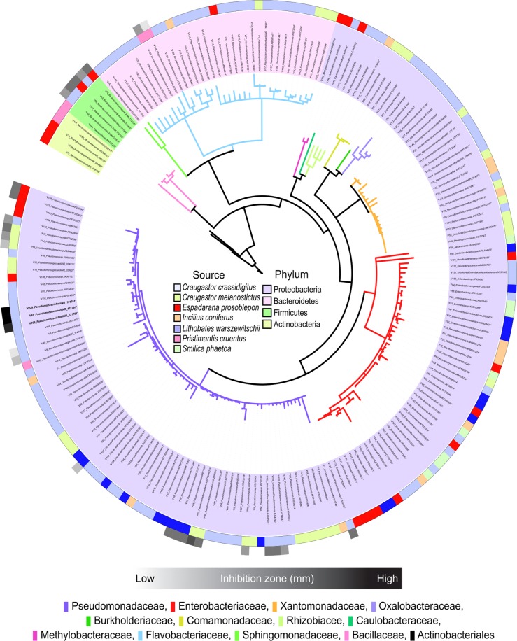
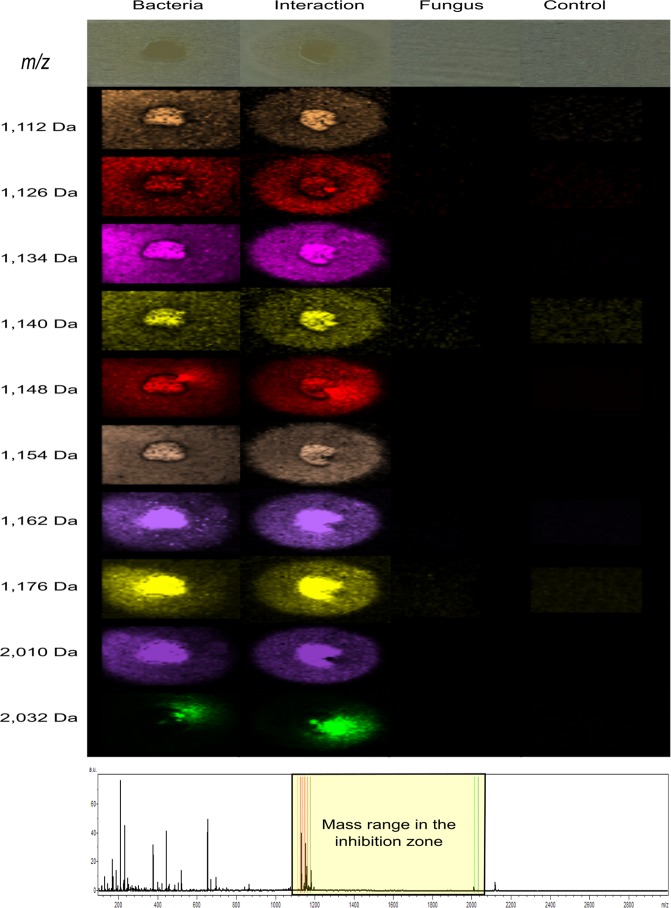
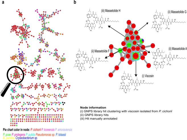
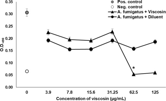
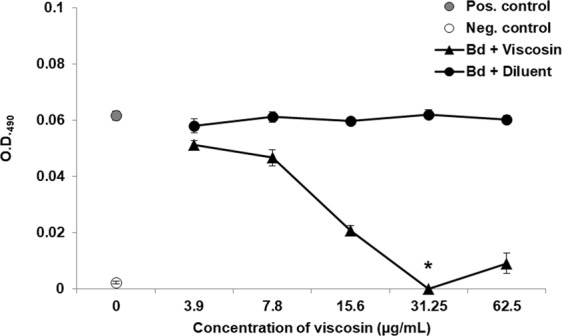
Amphibian populations worldwide have declined and in some cases become extinct due to chytridiomycosis, a pandemic disease caused by the fungus Batrachochytrium dendrobatidis; however, some species have survived these fungal epidemics. Previous studies have suggested that the resistance of these species is due to the presence of cutaneous bacteria producing antifungal metabolites. As our understanding of these metabolites is still limited, we assessed the potential of such compounds against human-relevant fungi such as Aspergillus. In this work we isolated 201 bacterial strains from fifteen samples belonging to seven frog species collected in the highlands of Panama and tested them against Aspergillus fumigatus. Among the 29 bacterial isolates that exhibited antifungal activity, Pseudomonas cichorii showed the greatest inhibition. To visualize the distribution of compounds and identify them in the inhibition zone produced by P. cichorii, we employed MALDI imaging mass spectrometry (MALDI IMS) and MS/MS molecular networking. We identified viscosin and massetolides A, F, G and H in the inhibition zone. Furthermore, viscosin was isolated and evaluated in vitro against A. fumigatus and B. dendrobatidis showing MIC values of 62.50 µg/mL and 31.25 µg/mL, respectively. This is the first report of cyclic depsipeptides with antifungal activity isolated from frog cutaneous bacteria.
Conflict of interest statement
The authors declare no competing interests.
Figures

Similar articles
-
Prodigiosin, Violacein, and Volatile Organic Compounds Produced by Widespread Cutaneous Bacteria of Amphibians Can Inhibit Two Batrachochytrium Fungal Pathogens.Microb Ecol. 2018 May;75(4):1049-1062. doi: 10.1007/s00248-017-1095-7. Epub 2017 Nov 9. Microb Ecol. 2018. PMID: 29119317
-
Cell Density Effects of Frog Skin Bacteria on Their Capacity to Inhibit Growth of the Chytrid Fungus, Batrachochytrium dendrobatidis.Microb Ecol. 2016 Jan;71(1):124-30. doi: 10.1007/s00248-015-0701-9. Epub 2015 Nov 12. Microb Ecol. 2016. PMID: 26563320
-
Phylogenetic distribution of symbiotic bacteria from Panamanian amphibians that inhibit growth of the lethal fungal pathogen Batrachochytrium dendrobatidis.Mol Ecol. 2015 Apr;24(7):1628-41. doi: 10.1111/mec.13135. Epub 2015 Mar 23. Mol Ecol. 2015. PMID: 25737297
-
Frog skin epithelium: electrolyte transport and chytridiomycosis.Int J Biochem Cell Biol. 2012 Mar;44(3):431-4. doi: 10.1016/j.biocel.2011.12.002. Epub 2011 Dec 13. Int J Biochem Cell Biol. 2012. PMID: 22182598 Free PMC article. Review.
-
Characterization of Aspergillus fumigatus mutants with reduced susceptibility to caspofungin.Med Mycol. 2005 May;43 Suppl 1:S299-305. doi: 10.1080/13693780400029023. Med Mycol. 2005. PMID: 16110824 Review.
Cited by
-
New Benthic Cyanobacteria from Guadeloupe Mangroves as Producers of Antimicrobials.Mar Drugs. 2019 Dec 23;18(1):16. doi: 10.3390/md18010016. Mar Drugs. 2019. PMID: 31878034 Free PMC article.
-
The Exploration of Microbial Natural Products and Metabolic Interaction Guided by Mass Spectrometry Imaging.Bioengineering (Basel). 2022 Nov 18;9(11):707. doi: 10.3390/bioengineering9110707. Bioengineering (Basel). 2022. PMID: 36421108 Free PMC article. Review.
-
Amphibian skin bacteria contain a wide repertoire of genes linked to their antifungal capacities.World J Microbiol Biotechnol. 2025 Feb 27;41(3):78. doi: 10.1007/s11274-025-04292-z. World J Microbiol Biotechnol. 2025. PMID: 40011297 Free PMC article.
-
Genome Mining, Microbial Interactions, and Molecular Networking Reveals New Dibromoalterochromides from Strains of Pseudoalteromonas of Coiba National Park-Panama.Mar Drugs. 2020 Sep 3;18(9):456. doi: 10.3390/md18090456. Mar Drugs. 2020. PMID: 32899199 Free PMC article.
-
Does regulation hold the key to optimizing lipopeptide production in Pseudomonas for biotechnology?Front Bioeng Biotechnol. 2024 Feb 27;12:1363183. doi: 10.3389/fbioe.2024.1363183. eCollection 2024. Front Bioeng Biotechnol. 2024. PMID: 38476965 Free PMC article. Review.
References
-
- Berger, L., Speare, R. & Hyatt, A. Chytrid fungi and amphibian declines: Overview, implications and future directions. Environ. Aust. Canberra 23–33 (1999).
Publication types
MeSH terms
Substances
Supplementary concepts
LinkOut - more resources
Full Text Sources
Research Materials
