. 2019 Feb 28;9(1):3135.
doi: 10.1038/s41598-019-40008-8.

Identification of P2Y receptors involved in oleamide-suppressing inflammatory responses in murine microglia and human dendritic cells

Affiliations

Identification of P2Y receptors involved in oleamide-suppressing inflammatory responses in murine microglia and human dendritic cells

Masahiro Kita et al. Sci Rep. .

Abstract

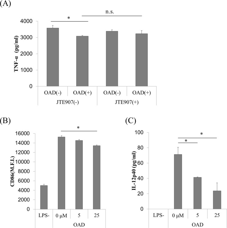
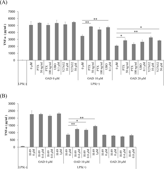
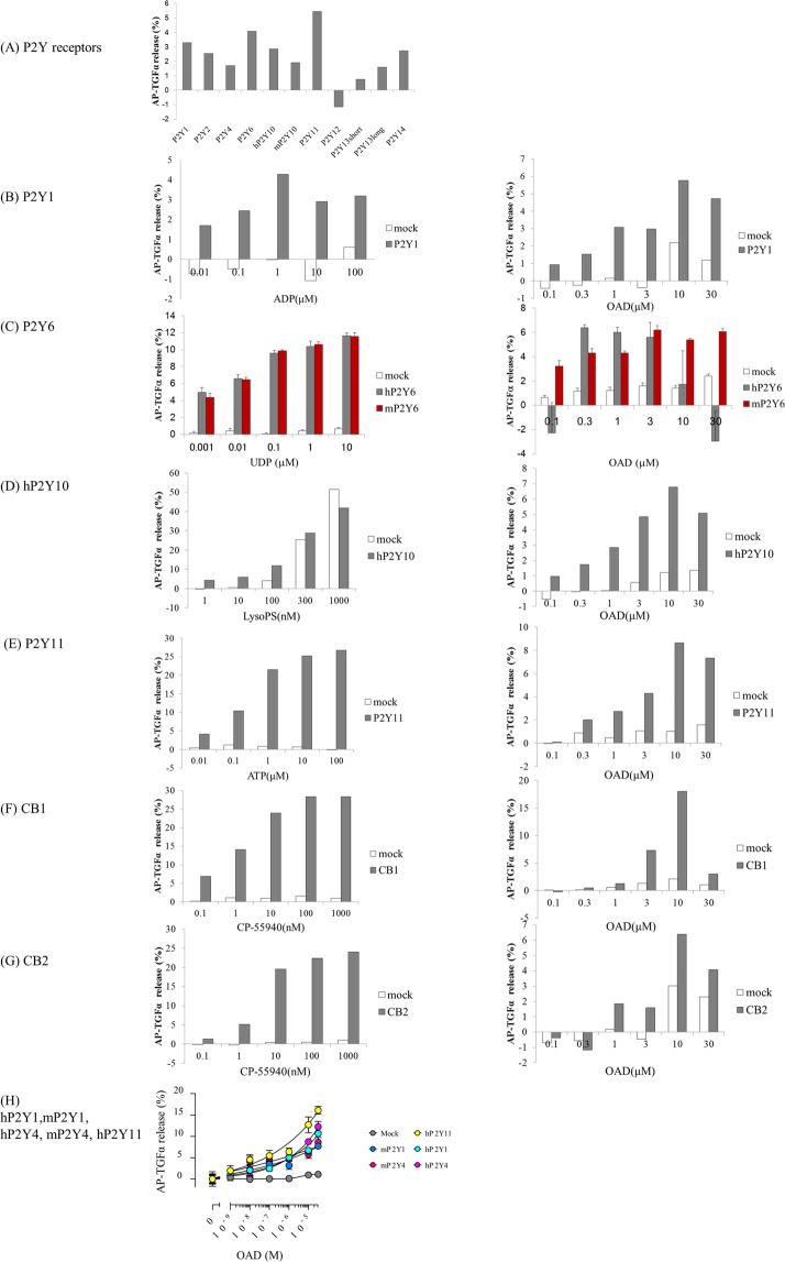
Microglia, a type of immune cell in the central nervous system, are involved in inflammation leading to neurodegenerative diseases. We previously identified oleamide from fermented dairy products as a neuroprotective compound suppressing microglial inflammation. Oleamide is an endocannabinoid and displays anti-inflammatory activity via the cannabinoid-2 (CB2) receptor; however, the mechanism underlying this anti-inflammatory activity has not been fully elucidated. Here, we found that the suppressive effect of oleamide on microglial tumor necrosis factor-α (TNF-α) production was canceled by inhibitors of G-protein-coupled receptor (GPCR) downstream signaling but not by a CB2 antagonist, suggesting that GPCRs other than CB2 are involved in the anti-inflammatory effects of oleamide. An extensive screen for GPCRs using a transforming growth factor-α shedding assay system identified P2Y1, P2Y4, P2Y6, P2Y10, and P2Y11 as candidates for the oleamide target. P2Y1 and P2Y10 agonists suppressed microglial TNF-α production, while a pan P2 receptor antagonist canceled the suppressive effect. Furthermore, we observed a relationship between the P2Y1 agonistic activities and the suppressive activities of oleamide and its analogs. Taken together, our results suggest that, in addition to CB2, P2Y type receptors are the potential targets of oleamide, and P2Y1 plays a role in the suppression of microglial inflammatory responses by oleamide. (200/200 words).


Conflict of interest statement

M.K. and Y.A. are employed by Kirin Co. Ltd. All other authors declare no competing interests.

Figures

Figure 1
Figure 1
The effects of OAD and JTE907 on inflammatory responses in murine microglia and human DCs. (A) Primary murine microglia were treated with 100 ng/ml of JTE907 for 1 h, followed by 5 μM OAD overnight. They were then stimulated by 5 ng/ml of LPS and 0.5 ng/ml of IFN-γ for 12 h. Extracellular TNF-α was measured by ELISA. (B,C) Human DCs were treated with OAD for 24 h, followed by 1 μg/ml of LPS, and 100 ng/ml of IFN-γ for 24 h. The expression of CD86 was then measured by FACS (B), and extracellular IL-12p40 was measured by ELISA (C). The data represents mean ± S.E.M. (n = 3) (*shows p < 0.05 tested by Tukey–Kramer’s test).
Figure 2
Figure 2
The effects of inhibitors of GPCR downstream signaling on the suppression of TNF-α production by OAD in murine microglia. (A,B) Primary mice microglia were treated with 50 or 100 ng/ml of PTX for 6 h (A), 0.1 μM UBO-QIC for 1 h (A), 10 or 50 μM Y27632 (A), or 0.4, 0.8, or 1 μM H-89 (Gs signal inhibitor) for 1 h (B), followed by OAD treatment overnight. They were then stimulated by 5 ng/ml of LPS and 0.5 ng/ml of IFN-γ for 12 h. Extracellular TNF-α was measured by ELISA. The data represents mean ± S.E.M. (A, n = 3; B, n = 5). (*p < 0.05, **p < 0.01 vs 0 μM control by Dunnett’s test).
Figure 3
Figure 3
Improvement in the TGF-α shedding response to OAD in HEK293 cells expressing P2Y1, P2Y4, P2Y6, P2Y10, P2Y11, CB1, and CB2. (A) Summary of the results of the TGF-α shedding response in cells transfected with P2Y receptors during the screening series. Mock transfected cells (pCAGGS empty vector) (white bar) (BH) or HEK 293 cells expressing GPCRs (left) (P2Y1 (B,H), human P2Y6 (C, dark bar), mouse P2Y6 (B, red bar), P2Y10 (D), P2Y11 (E,H), CB1 (F), CB2 (G), and human and mouse P2Y4 (H)) were treated with OAD (right) or each receptor agonist (left) (ADP (B), UDP (C), LysoPS (D), ATP (E), CP-55940 (F,G)). The TGF-α shedding response was then calculated. Data represent the mean (n = 2) (A,B, DF), mean ± S.E.M (n = 4) (C), or mean ± S.E.M (n = 3) (H).
Figure 4
Figure 4
The effects of GPCR agonists and antagonist on microglial TNF-α production. (A,B) Primary mice microglia were treated with CP-55940 (A), or LysoPS, a P2Y10-selective agonist, or MRS2365 (B) overnight. The data represent mean ± S.E.M (n = 3–6). (*p < 0.05 vs 0 μM control by Williams’s test in each agonist group.) (C) Cells were treated with different concentrations of suramin for 30 min, followed by 20 μM OAD. Data represents mean ± S.E.M. (n = 3). (*p < 0.05, **p < 0.01 vs 0 μM control by two-tail Dunnett’s test).
Figure 5
Figure 5
Comparison of agonistic activity and TNF-α suppressing activity between OAD and its analogs. (A) Cells expressing human and murine P2Y1 and P2Y4, and human P2Y11 were treated with OAD, tOAD, oleic acid, OEtA, PEA, or OEA, and AP-TGFα release was measured. (B) Primary microglia were treated with OAD and its analog derived from oleic acid (B, upper), or PEA (B, lower) overnight, followed by LPS stimulation. Bars with a different letter were statistically different by Turkey–Kramer’s test. Statistical analysis was performed between a control 0 μM value and each concentration. The data represent mean ± S.E.M (n = 3).

References

    1. Bolos M, Perea JR, Avila J. Alzheimer’s disease as an inflammatory disease. Biomol Concepts. 2017;8(1):37–43. doi: 10.1515/bmc-2016-0029. - DOI - PubMed
    1. Ramirez AI, et al. The Role of Microglia in Retinal Neurodegeneration: Alzheimer’s Disease, Parkinson, and Glaucoma. Front Aging Neurosci. 2017;9:214. doi: 10.3389/fnagi.2017.00214. - DOI - PMC - PubMed
    1. Chen WW, Zhang X, Huang WJ. Role of neuroinflammation in neurodegenerative diseases (Review) Mol Med Rep. 2016;13(4):3391–6. doi: 10.3892/mmr.2016.4948. - DOI - PMC - PubMed
    1. Salter MW, Stevens B. Microglia emerge as central players in brain disease. Nat Med. 2017;23(9):1018–1027. doi: 10.1038/nm.4397. - DOI - PubMed
    1. G. Olmos, J. Llado, Tumor necrosis factor alpha: a link between neuroinflammation and excitotoxicity. Mediators Inflamm. 861231 (2014). - PMC - PubMed