. 2019 Feb 28;9(1):3128.
doi: 10.1038/s41598-019-39997-3.

Inhibitory effects of polysorbate 80 on MRSA biofilm formed on different substrates including dermal tissue

Affiliations

Inhibitory effects of polysorbate 80 on MRSA biofilm formed on different substrates including dermal tissue

Yutaka Ueda et al. Sci Rep. .

Abstract

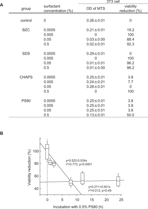
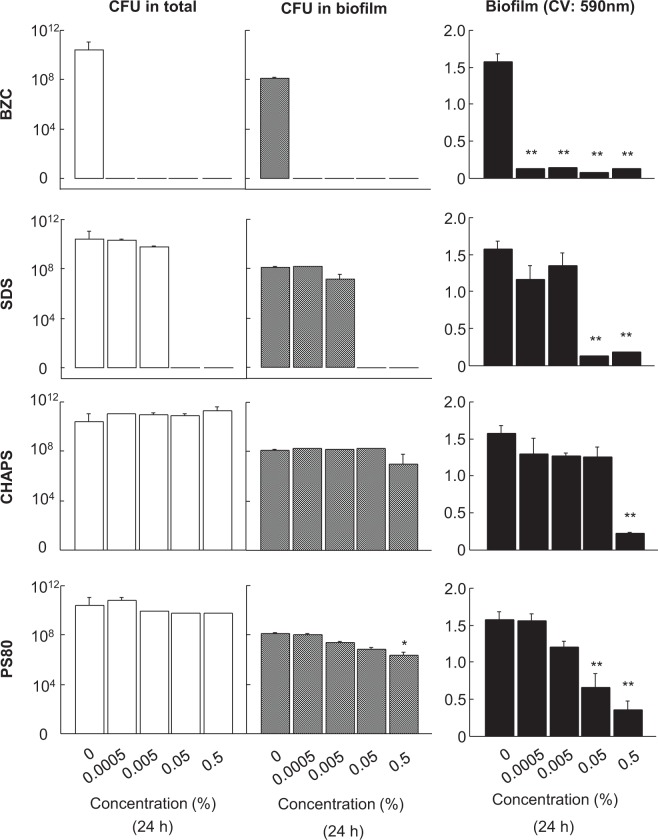
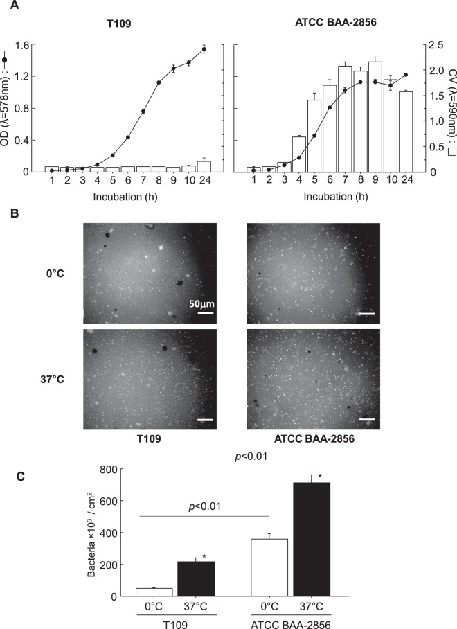
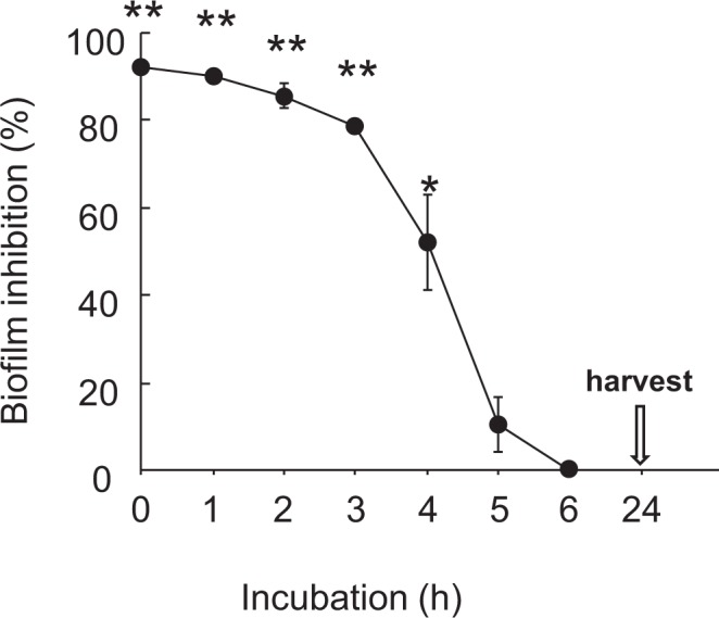
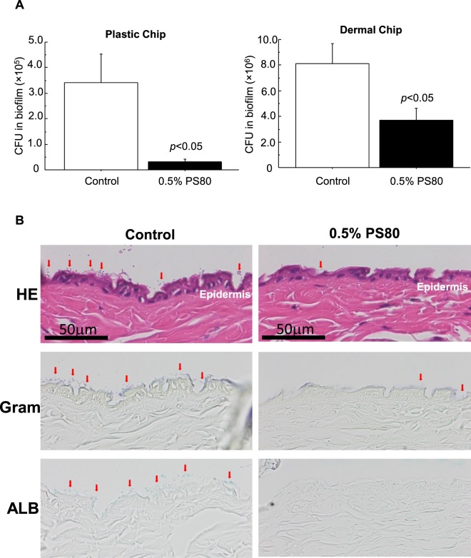
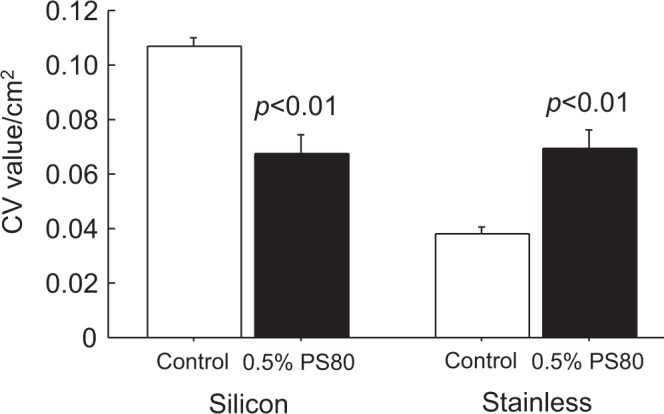
Methicillin-resistant Staphylococcus aureus (MRSA) forms biofilms on necrotic tissues and medical devices, and causes persistent infections. Surfactants act on biofilms, but their mode of action is still unknown. If used in the clinic, cytotoxicity in tissues should be minimized. In this study, we investigated the inhibitory effect of four different surfactants on MRSA biofilm formation, and found that a nonionic surfactant, polysorbate 80 (PS80), was the most suitable. The biofilm inhibitory effects resulted from the inhibition of bacterial adhesion to substrates rather than biofilm disruption, and the effective dose was less cytotoxic for 3T3 fibroblasts. However, the effects were substrate-dependent: positive for plastic, silicon, and dermal tissues, but negative for stainless-steel. These results indicate that PS80 is effective for prevention of biofilms formed by MRSA on tissues and foreign bodies. Therefore, PS80 could be used in medical practice as a washing solution for wounds and/or pretreatment of indwelling catheters.


Conflict of interest statement

The authors declare no competing interests.

Figures

Figure 1
Figure 1
Effect of different surfactants on 3T3 fibroblast viability. (A) Different types of surfactants at concentrations up to 0.5% were added to 3T3 cultures in 96 well plates. After incubation for 24 h at 37 °C, MTS viability assay was performed. Values are viability reduction rates (%) against the vehicle control. Although all surfactants in this study were used at less than MTS50 cytotoxic doses, PS80 showed the mildest effect in the range. Data are presented as mean ± SE. Sample number/group = 8, duplicated. (B) Using 0.5% PS80 in the culture, effects of exposure time on 3T3 cell viability were examined. Cell viability was reduced to about 50% in the first 8 h of incubation, however, it was not further aggravated during 24 h of incubation. Sample number/group = 16, duplicated.
Figure 2
Figure 2
Effects of different surfactants on CFU and biofilm formation values of ATCC BAA-2856 cultured in plastic tubes. Different types of surfactants at concentrations up to 0.5% were added to plastic tubes containing a 1000-fold diluted bacterial solution of ATCC BAA-2856, a biofilm forming MRSA at OD = 0.57, and was incubated for 24 h at 37 °C. After incubation, total bacteria in the tube and in the biofilm formed on the tube surface were assessed using CFU analysis. Biofilm mass was also measured by CV staining. Data are presented as mean ± SE, *p < 0.05, **p < 0.01 vs. vehicle control. Sample number/group = 3, triplicated.
Figure 3
Figure 3
Time courses of bacterial growth and biofilm formation for low and high biofilm formers in plastic tubes and bacterial attachment on a plastic substrate at 0 °C and 37 °C. (A) T109 and ATCC BAA-2856 were used for the study. Bacteria were sparsely seeded in plastic tubes, and cultured for 24 h. Bacterial growth was assessed as medial OD values (solid line), and biofilm mass was measured with CV staining (open column). The results clearly showed that the bacteria were low and high biofilm-forming MRSA, respectively. Data: mean ± SE. Sample number/group = 3, quadruplicated. (B) Plastic chips (1 × 1 cm pieces of OHP sheet) were immersed in confluent bacterial solutions, and incubated at 0 °C and 37 °C for 1 h. After incubation, bacteria adhered on the surface were visualized using FITC-labeled S. aureus antibody. Bar = 50 μm. (C) Bacteria adhered on plastic chips were counted using the morphometric method as described in the Materials and Methods. Data are presented as bacteria number/cm2 area, mean ± SE, *p < 0.01 vs. 0 °C. Sample number/group ≥9, duplicated.
Figure 4
Figure 4
Inhibitory effects of 0.5% PS80 addition at different time points on biofilm formation during 24 h incubation. During the 24 h biofilm assay starting from 1000-fold diluted ATCC BAA-2856 bacterial solution at OD = 0.57, 0.5% PS80 was added at 0, 1, 2, 3, 4, 5, and 6 h of incubation. At the end of the study, the biofilm mass formed on the tube wall surface was measured with CV stain. Data are presented as mean ± SE. Sample number/group = 3, triplicated.
Figure 5
Figure 5
Inhibitory effects of 0.5% PS80 on bacterial attachment to dermal and plastic chips. (A) Plastic chips (1 × 1 cm pieces of OHP sheet) and dermal chip (1 × 1 cm pieces of mouse skin) were immersed in confluent bacterial solutions with/without 0.5% PS80, and incubated at 37 °C for 1 h. After incubation, bacteria adhered on the chips were assessed by CFU analysis. Data are presented as mean ± SE. Sample number/group = 5, duplicated. (B) Serial sections of dermal chips were subjected to HE, Gram and ALB stainings after incubation in confluent bacterial solutions. Gram-positive bacteria attached on the surface show a fine spherical structure (red arrows) and ALB-positive EPS containing biofilm (red arrows). Addition of 0.5% PS80 apparently decreased the number of attached bacteria accompanied with biofilm on the dermal tissue.
Figure 6
Figure 6
Inhibitory effects of 0.5% PS80 on biofilm formation with silicon and stainless steel substrates. Diluted ATCC BAA-2856 bacterial solutions (1000-fold from OD = 0.57) with/without 0.5% PS80 were added to silicon-coated tubes and incubated for 24 h at 37 °C. Stainless steel sheets cut to 3 × 3 cm were also immersed in bacterial solutions with/without 0.5% PS80 for 24 h at 37 °C. After incubation, the biofilm mass formed on the silicon substrate and stainless steel substrate was measured by CV staining. Data are presented as CV values/cm2, mean ± SE. Sample number/group = 10, triplicated.

References

    1. DeLeo FR, Chambers HF. Reemergence of antibiotic-resistant Staphylococcus aureus in the genomics era. J Clin Invest. 2009;119:2464–2474. doi: 10.1172/JCI38226. - DOI - PMC - PubMed
    1. Otto M. Staphylococcal infections: mechanisms of biofilm maturation and detachment as critical determinants of pathogenicity. Annu Rev Med. 2013;64:175–188. doi: 10.1146/annurev-med-042711-140023. - DOI - PubMed
    1. Kiedrowski MR, Horswill AR. New approaches for treating staphylococcal biofilm infections. Ann N Y Acad Sci. 2011;1241:104–121. doi: 10.1111/j.1749-6632.2011.06281.x. - DOI - PubMed
    1. Utsui Y, Yokota T. Role of an altered penicillin-binding protein in methicillin- and cephem-resistant Staphylococcus aureus. Antimicrob Agents Chemother. 1985;28:397–403. doi: 10.1128/AAC.28.3.397. - DOI - PMC - PubMed
    1. Evans RC, Holmes CJ. Effect of vancomycin hydrochloride on Staphylococcus epidermidis biofilm associated with silicone elastomer. Antimicrob Agents Chemother. 1987;31:889–894. doi: 10.1128/AAC.31.6.889. - DOI - PMC - PubMed

Publication types

MeSH terms