

Adipose‑derived mesenchymal stem cells exhibit tumor tropism and promote tumorsphere formation of breast cancer cells
- PMID: 30816504
- PMCID: PMC6412463
- DOI: 10.3892/or.2019.7018
Adipose‑derived mesenchymal stem cells exhibit tumor tropism and promote tumorsphere formation of breast cancer cells
Abstract
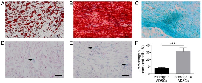
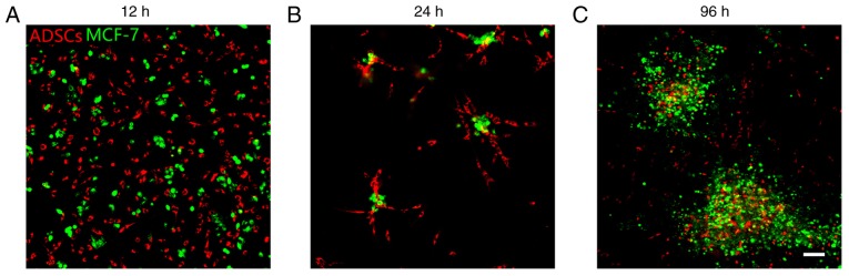
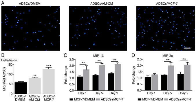
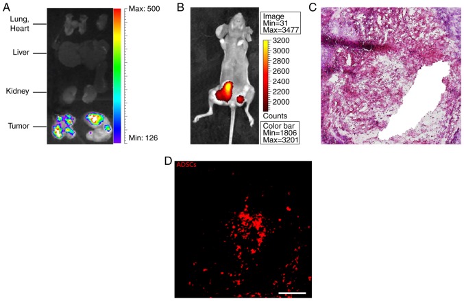
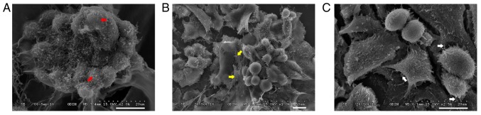
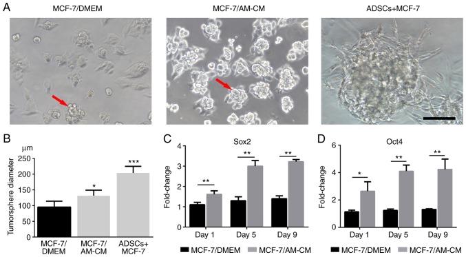
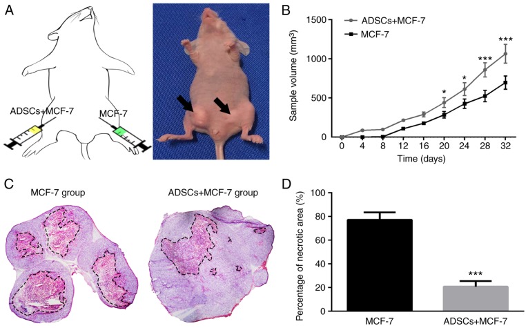
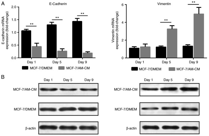
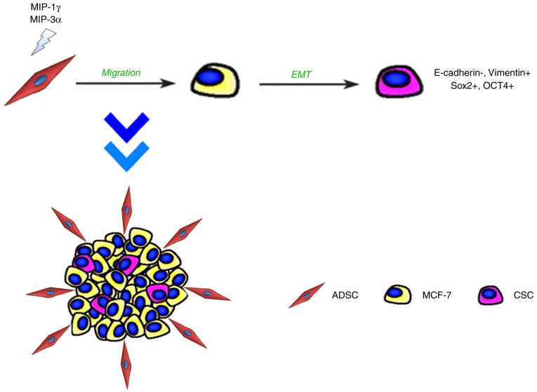
Mesenchymal stem cells reportedly have a marked effect on tumor growth or suppression. However, it remains uncertain whether adipose‑derived mesenchymal stem cells (ADSCs) from grafted fat can contribute to breast cancer growth and recurrence. In the present study, interactions between ADSCs and MCF‑7 breast cancer cells were evaluated in a Matrigel co‑culture system and in an in vivo nude mouse model. Results suggested that MCF‑7 cells exerted tumor tropism effects on ADSCs and this may be regulated by chemokines, such as the macrophage inflammatory protein (MIP)‑1δ and MIP‑3α. Additionally, ADSCs significantly induced tumorsphere formation in vitro and promoted tumorigenicity in vivo. RT‑qPCR analysis indicated that tumorsphere formation by MCF‑7 cells was associated with the induction of stem‑like properties, which was mediated by epithelial‑-mesenchymal transition. Together, the present findings indicated that ADSCs exhibit tropism and induce tumorsphere formation of MCF‑7 cells.
Figures
Similar articles
-
Human adipose‑derived mesenchymal stem cells promote breast cancer MCF7 cell epithelial‑mesenchymal transition by cross interacting with the TGF‑β/Smad and PI3K/AKT signaling pathways.Mol Med Rep. 2019 Jan;19(1):177-186. doi: 10.3892/mmr.2018.9664. Epub 2018 Nov 19. Mol Med Rep. 2019. PMID: 30483746 Free PMC article.
-
The impact of human adipose tissue-derived stem cells on breast cancer cells: implications for cell-assisted lipotransfers in breast reconstruction.Stem Cell Res Ther. 2017 May 25;8(1):121. doi: 10.1186/s13287-017-0579-1. Stem Cell Res Ther. 2017. PMID: 28545495 Free PMC article.
-
The metastatic phenotype shift toward myofibroblast of adipose-derived mesenchymal stem cells promotes ovarian cancer progression.Carcinogenesis. 2020 Apr 22;41(2):182-193. doi: 10.1093/carcin/bgz083. Carcinogenesis. 2020. PMID: 31046126
-
Human Adipose-Derived Mesenchymal Stem Cell-Secreted CXCL1 and CXCL8 Facilitate Breast Tumor Growth By Promoting Angiogenesis.Stem Cells. 2017 Sep;35(9):2060-2070. doi: 10.1002/stem.2643. Epub 2017 Jun 5. Stem Cells. 2017. PMID: 28514506
-
Safety of adipose-derived cell (stromal vascular fraction - SVF) augmentation for surgical breast reconstruction in cancer patients.Adv Clin Exp Med. 2018 Aug;27(8):1085-1090. doi: 10.17219/acem/70798. Adv Clin Exp Med. 2018. PMID: 29989681
Cited by
-
Dual impacts of mesenchymal stem cell-derived exosomes on cancer cells: unravelling complex interactions.J Cell Commun Signal. 2023 Dec;17(4):1229-1247. doi: 10.1007/s12079-023-00794-3. Epub 2023 Nov 16. J Cell Commun Signal. 2023. PMID: 37973719 Free PMC article. Review.
-
Therapeutic Effects of Cold Atmospheric Plasma on Solid Tumor.Front Med (Lausanne). 2022 May 13;9:884887. doi: 10.3389/fmed.2022.884887. eCollection 2022. Front Med (Lausanne). 2022. PMID: 35646968 Free PMC article. Review.
-
The Targeted Impact of Flavones on Obesity-Induced Inflammation and the Potential Synergistic Role in Cancer and the Gut Microbiota.Molecules. 2020 May 27;25(11):2477. doi: 10.3390/molecules25112477. Molecules. 2020. PMID: 32471061 Free PMC article. Review.
-
Visfatin Mediates Malignant Behaviors through Adipose-Derived Stem Cells Intermediary in Breast Cancer.Cancers (Basel). 2019 Dec 20;12(1):29. doi: 10.3390/cancers12010029. Cancers (Basel). 2019. PMID: 31861872 Free PMC article.
-
Adipose-derived stem cells promote the proliferation, migration, and invasion of oral squamous cell carcinoma cells by activating the Wnt/planar cell polarity signaling pathway.Transl Cancer Res. 2022 Feb;11(2):306-315. doi: 10.21037/tcr-21-1637. Transl Cancer Res. 2022. PMID: 35281413 Free PMC article.
References
-
- Nelson HD, Zakher B, Cantor A, Fu R, Griffin J, O'Meara ES, Buist DS, Kerlikowske K, van Ravesteyn NT, Trentham-Dietz A, et al. Risk factors for breast cancer for women aged 40 to 49 years: A systematic review and meta-analysis. Ann Intern Med. 2012;156:635–648. doi: 10.7326/0003-4819-156-9-201205010-00006. - DOI - PMC - PubMed
-
- Munhoz AM, Montag E, Filassi JR, Gemperli R. Current approaches to managing partial breast defects: The role of conservative breast surgery reconstruction. Anticancer Res. 2014;34:1099–1114. - PubMed
-
- Longaker MT, Aston SJ, Baker DC, Rohrich RJ. Fat Transfer in 2014: What we do not know. Plast Reconstr Surg. 2014;133:1305–1307. - PubMed
MeSH terms
LinkOut - more resources
Full Text Sources
Medical
