. 2019 Apr;19(4):3009-3020.
doi: 10.3892/mmr.2019.9959. Epub 2019 Feb 15.

Neuroprotection of hydroxysafflor yellow A in experimental cerebral ischemia/reperfusion injury via metabolic inhibition of phenylalanine and mitochondrial biogenesis

Affiliations

Neuroprotection of hydroxysafflor yellow A in experimental cerebral ischemia/reperfusion injury via metabolic inhibition of phenylalanine and mitochondrial biogenesis

Suning Chen et al. Mol Med Rep. 2019 Apr.

Abstract

Stroke is the second most frequent cause of mortality, resulting in a huge societal burden worldwide. Timely reperfusion is the most effective therapy; however, it is difficult to prevent ischemia/reperfusion (I/R) injury. In traditional Chinese medicine, hydroxysafflor yellow A (HSYA) has been widely used for the treatment of cerebrovascular disease and as a protective therapy against I/R injury. Evidence has demonstrated that HSYA could reduce the levels of reactive oxygen species and suppress cellular apoptosis; however, whether HSYA alters the metabolic profile as its underlying mechanism for neuroprotection remains unknown. In the present study, using a metabolomic screening, phenylalanine was identified to significantly increase in an experimental model of mouse cerebral I/R injury. Notably, western blotting and qPCR analysis were conducted to test the expression level of apoptosis‑associated factors, and HSYA was identified to be able to protect neuronal cells by reducing phenylalanine level associated with I/R injury. Additionally, these findings were confirmed in primary mouse neurons and PC12 cells exposed to oxygen and glucose deprivation/reoxygenation (OGD/R) stress. Of note, HSYA was observed to regulate the mRNA expression of key metabolic enzymes, phenylalanine hydroxylase, tyrosine aminotransferase and aspartate aminotransferase, which are responsible for phenylalanine metabolism. Furthermore, by performing mitochondrial labeling and JC‑1 fluorescence assay, HSYA was identified to promote mitochondrial function and biogenesis suppressed by OGD/R. The findings of the present study demonstrated that I/R injury could increase the levels of phenylalanine, and HSYA may inhibit phenylalanine synthesis to enhance mitochondrial function and biogenesis for neuroprotection. The present study proposed a novel metabolite biomarker for cerebral I/R injury and the evaluated the efficacy of HSYA as a potential therapeutic treatment I/R injury.


Figures

Figure 1.
Figure 1.
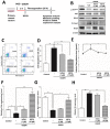
HSYA alleviates neurological deficits and reduces the infarct size induced by cerebral I/R. (A) Experimental scheme for the experimental mice cerebral I/R. Briefly, mice received ischemia via middle cerebral artery occlusion for 2 h and reperfusion for 3 days, with or without HSYA (5 or 20 mg/kg) treatment. (B) Neurological behavior deficit evaluation. A scale of 0–4 points was used to evaluate the neurological deficits. The values are expressed as the mean ± standard error of the mean of 6 mice in each group. I/R significantly increased deficit score, but was reduced following treatment with HSYA. (C) HSYA reduced the infarct zone. TTC staining was used to monitor the infarct area (white). I/R resulted in an infarct zone; HSYA decreased the size of the area. Scale bar=5 mm. (D) Results of statistical analysis of the TTC staining zone with three mice of each group (n=3). (E) Western blotting was conducted to analyze the expression of proliferation and apoptosis-associated molecules with antibodies against p-Akt, β-catenin, BCL2, cleaved caspase-3 and neuron marker NeuN. β-actin was the internal control. (F) Band intensity quantification of NeuN. *P<0.05, **P<0.01. Akt, protein kinase B; BCL2, B-cell lymphoma 2; c-Casp3, cleaved caspase 3; HYSA, hydroxysafflor yellow A; I/R, ischemia/reperfusion; NeuN, neuronal nuclei; p, phosphorylated; TTC, 2, 3, 5-triphenyltetrazolium chloride.
Figure 2.
Figure 2.
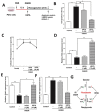
HSYA regulates the levels of phenylalanine and the metabolic enzymes expression caused by cerebral I/R in vivo. (A) I/R stress increased the plasma levels of phenylalanine, which was reversed by HSYA. (B) Transformation flux of phenylalanine. Phenylalanine can be metabolized to phenylpyruvate and tyrosine via GOT1 and TAT, and PAH, respectively. HSYA reduced the levels of phenylalanine by regulating the expression of key metabolic enzymes. mRNA expression and relative quantification of (C) Pah, (D) Got1 and (E) Tat. The data are expressed as the mean ± standard error of the mean. *P<0.05, **P<0.01. GOT1, aspartate aminotransferase; HYSA, hydroxysafflor yellow A; I/R, ischemia/reperfusion; PAH, phenylalanine hydroxylase; TAT, tyrosine aminotransferase.
Figure 3.
Figure 3.
HSYA regulates the levels of phenylalanine in primary mouse neuronal cells with OGD/R stress. (A) Experimental scheme for primary mouse neurons with OGD/R treatment. (B) Analysis of p-Akt, BCL2, cleaved caspase-3 and NeuN levels by means of western blotting in primary mouse neuron with OGD/R treatment. (C) Analysis of apoptosis via flow cytometry. (D) Quantification of apoptosis. (E) Quantification of phenylalanine levels in the control and OGD/R groups treated with or without HSYA. HSYA regulates the expression of key metabolic enzymes in primary mouse neuronal cells. mRNA expression and relative quantification of (F) Pah, (G) Got1 and (H) Tat. The data are expressed as the mean ± standard error of the mean. *P<0.05, **P<0.01. Akt, protein kinase B; BCL2, B-cell lymphoma 2; c-Casp3, cleaved caspase 3; GOT1, aspartate aminotransferase; HYSA, hydroxysafflor yellow A; PAH, phenylalanine hydroxylase; NeuN, neuronal nuclei; OGD/R, oxygen and glucose deprivation/reoxygenation; p, phosphorylated; TAT, tyrosine aminotransferase.
Figure 4.
Figure 4.
HSYA suppresses the apoptosis and alters the expression of enzymes associated with the metabolism of phenylalanine in PC12 cells with OGD/R stress. (A) Experimental scheme for PC12 cells exposed to OGD/R. (B) Apoptosis analysis and quantification in each group via flow cytometry. (C) Quantification of phenylalanine levels in the control and OGD/R groups treated with or without HSYA. HSYA regulated the expression of key metabolic enzymes of phenylalanine in PC12 cells. Relative quantification of the mRNA expression of (D) Pah, (E) Got1 and (F) Tat. (G) Summarization of HSYA regulating metabolic enzymes expression to reduce the levels of phenylalanine. All data are expressed as the mean ± standard error of the mean. *P<0.05, **P<0.01. Got1, aspartate aminotransferase; HYSA, hydroxysafflor yellow A; Pah, phenylalanine hydroxylase; OGD/R, oxygen and glucose deprivation/reoxygenation; p, phosphorylated; Tat, tyrosine aminotransferase.
Figure 5.
Figure 5.
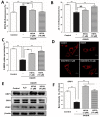
HSYA promotes mitochondrial function and biogenesis in PC12 cells exposed to OGD/R stress. (A) DCFH-DA analysis in PC12 cells with OGD/R stress and HSYA treatment as indicated. (B) JC-1 red fluorescence assay in PC12 cells with OGD/R stress and HSYA treatment. (C) Glucose uptake ability in PC12 cells exposed to OGD/R stress and HSYA as detected by 2-NBDG labelling. (D) MitoTracker Red fluorescence in PC12 cells was detected with a confocal immunofluorescence microscope. Mitochondrial shape was compared in each group. Scale bar=50 µm. (E) Western blotting analysis of mitochondrial fission protein DRP1, fusion protein OPA1 and mitochondrial marker VDAC. β-actin was the internal control. (F) Band intensity quantification of DRP1 in (E). All data are expressed as the mean ± standard error of the mean. **P<0.01. 2-NBDG, 2-[N-(7-nitrobenz-2-oxa-1,3-diazol-4-yl)amino]-2-deoxyglucose; DCFH-DA, dichloro-dihydro-fluorescein diacetate; DRP1, dynamin-1-like protein; HYSA, hydroxysafflor yellow A; OPA1, dynamin-like GTPase; NS, not significant; VDAC, voltage-dependent anion-selective channel 1; OGD/R, oxygen and glucose deprivation/reoxygenation.
Figure 6.
Figure 6.
HSYA protects mitochondrial function suppressed by PHE in PC12 cells. (A) MTT assay in PC12 cells with PHE treatment for 2 and 4 days with increased concentration (0, 0.5, 1, 2, 4, 8, 16 and 32 mM). (B) JC-1 red fluorescence assay in PC12 cells with 8 mM PHE and HSYA (1 and 10 µM) treatment as indicated. (C) Western blotting analysis of mitochondrial fission protein DRP1 and Fis1, and fusion protein MFN2. β-actin was the internal control. (D) Band intensity quantification of DRP1. All data are expressed as the mean ± standard error of the mean. *P<0.05, **P<0.01. DRP1, dynamin-1-like protein; Fis1, mitochondrial fission 1 protein; HYSA, hydroxysafflor yellow A; MFN2, mitofusin-2; OGD/R, oxygen and glucose deprivation/reoxygenation; PHE, phenylalanine.
Figure 7.
Figure 7.
Diagram of HSYA regulating the levels of phenylalanine, mitochondrial function and biogenesis for neuroprotection. I/R injury caused increased ROS and phenylalanine levels to induce neuronal cell apoptosis. HSYA could reduce the production of ROS and phenylalanine by regulating the expression of key metabolic enzymes, including PAH, TAT and GOT1. In addition, HSYA could promote mitochondrial function and biogenesis by upregulating mitochondria fission protein DRP1. GOT1, aspartate aminotransferase; GSH, glutathione; HYSA, hydroxysafflor yellow A; PAH, phenylalanine hydroxylase; ROS, reactive oxygen species; SOD, superoxide dismutase.

References

    1. Chu SF, Zhang Z, Zhang W, Zhang MJ, Gao Y, Han N, Zuo W, Huang HY, Chen NH. Upregulating the expression of Survivin-HBXIP complex contributes to the protective role of IMM-H004 in transient global cerebral Ischemia/reperfusion. Mol Neurobiol. 2017;54:524–540. doi: 10.1007/s12035-015-9673-5. - DOI - PubMed
    1. Sarami Foroshani M, Sobhani ZS, Mohammadi MT, Aryafar M. Fullerenol nanoparticles decrease blood-brain barrier interruption and brain edema during cerebral ischemia-reperfusion injury probably by reduction of interleukin-6 and matrix metalloproteinase-9 transcription. J Stroke Cerebrovasc Dis. 2018;27:3053–3065. doi: 10.1016/j.jstrokecerebrovasdis.2018.06.042. - DOI - PubMed
    1. Wiklund L, Patnaik R, Sharma A, Miclescu A, Sharma HS. Cerebral tissue oxidative ischemia-reperfusion injury in connection with experimental cardiac arrest and cardiopulmonary resuscitation: Effect of mild hypothermia and methylene blue. Mol Neurobiol. 2018;55:115–121. doi: 10.1007/s12035-017-0723-z. - DOI - PMC - PubMed
    1. Gong L, Tang Y, An R, Lin M, Chen L, Du J. RTN1-C mediates cerebral ischemia/reperfusion injury via ER stress and mitochondria-associated apoptosis pathways. Cell Death Dis. 2017;8:e3080. doi: 10.1038/cddis.2017.465. - DOI - PMC - PubMed
    1. Li Y, Wang M, Wang S. Effect of inhibiting mitochondrial fission on energy metabolism in rat hippocampal neurons during ischemia/reperfusion injury. Neurol Res. 2016:1–8. (Epub ahead of print) - PubMed

MeSH terms