. 2019 Apr;19(4):3139-3147.
doi: 10.3892/mmr.2019.9963. Epub 2019 Feb 18.

Colony‑stimulating factor 1 receptor inhibition blocks macrophage infiltration and endometrial cancer cell proliferation

Affiliations

Colony‑stimulating factor 1 receptor inhibition blocks macrophage infiltration and endometrial cancer cell proliferation

Fu Hua et al. Mol Med Rep. 2019 Apr.

Abstract

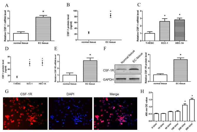
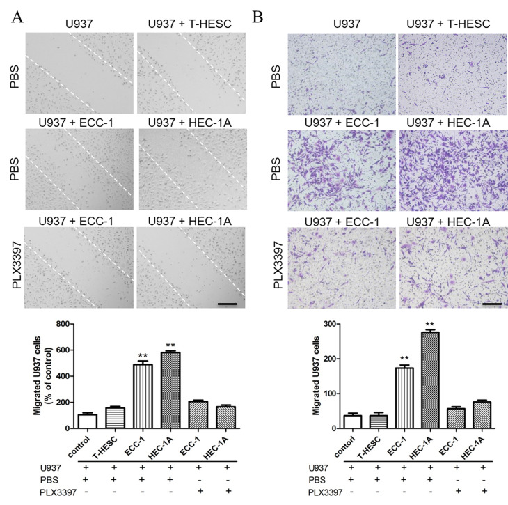
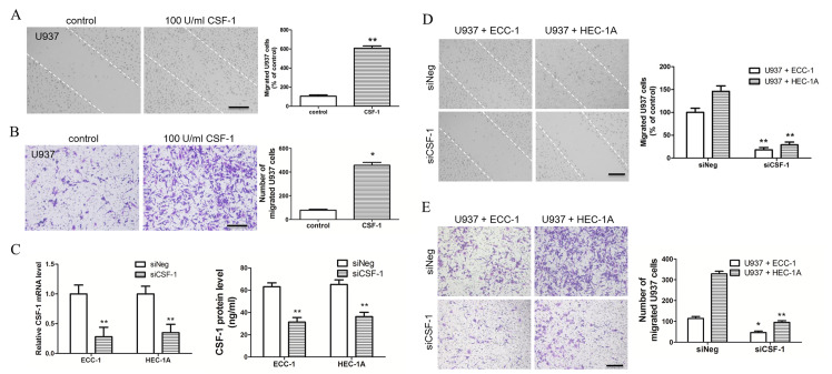
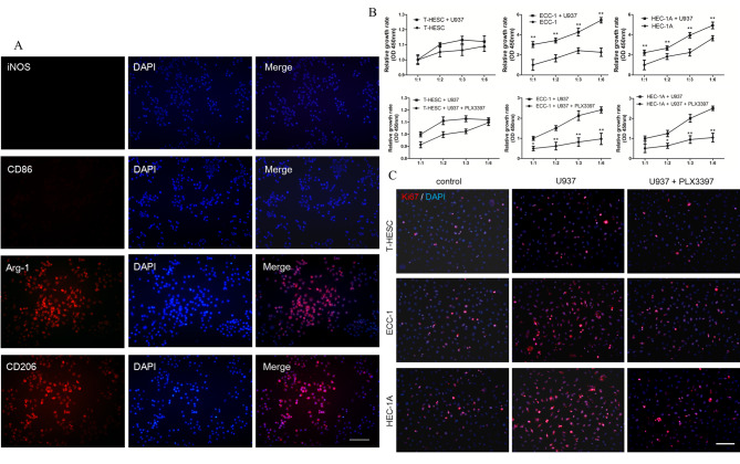
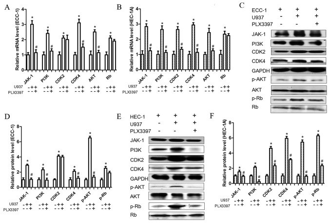
Tumor‑associated macrophages (TAMs) promote the progression of endometrial cancer (EC), but the mechanism of TAM in EC cell proliferation remains unclear. It was found that colony stimulating factor (CSF)‑1 and CSF‑1 receptor (CSF‑1R) were highly expressed in EC tissues of patients and two EC cell lines (ECC‑1 and HEC‑1A). Using wound‑healing and chemotactic migration assays to evaluate the role of EC cells in the induction of macrophage migration, it was found that the supernatant of EC cells promoted macrophage cell line (U937) migration; however, the migration capacity of U937 weakened when CSF‑1R was blocked. Subsequently, inhibition of CSF‑1 expression in EC cells also restrained U937 migration. Additionally, blocking CSF‑1R by PLX3397 treatment in U937 cells inhibited EC cell proliferation in a co‑culture system by inhibiting the expression of proliferation‑associated proteins (Janus kinase‑1, phosphoinositide 3‑kinase, AKT, cyclin kinase 2, 4 and retinoblastoma‑associated protein). Together, these results demonstrated that CSF‑1 secreted by EC cells promoted macrophage migration; similarly, CSF‑1‑stimulated macrophages promoted EC cell proliferation. These results suggested that the interaction between CSF‑1 and its receptor served an important role in promoting macrophage infiltration and progression of EC.


Figures

Figure 1.
Figure 1.
Expression levels of CSF-1 and CSF-1R in EC cells and macrophages. (A) mRNA and (B) protein expression levels of CSF-1 in endometrial tissues. (C) mRNA and (D) protein expression levels of CSF-1 in normal endometrial cells (T-HESC) and EC cells (ECC-1 and HEC-1A). (E) mRNA and (F) protein expression levels of CSF-1R in endometrial tissues. (G) Immunofluorescence detected CSF-1R expression levels in U937 cell lines. (H) Cell counting kit-8 assay detected the proliferation of U937 cells stimulated by different concentrations of CSF-1. Data are presented as the mean ± standard deviation from 10 (tissues) or 5 (cells) independent experiments; *P<0.05 vs. Control. Scale bar, 100 µm. CSF, colony-stimulating factor; CSF-1R, colony-stimulating factor 1 receptor; EC, endometrial cancer; OD, optical density.
Figure 2.
Figure 2.
CSF-1R inhibition blocks U937 cell migration. (A) Wound-healing assay detected U937 cell migration. (B) Chemotactic migration assay detected U937 cells migration. Data are presented as the mean ± standard deviation from 5 independent experiments; **P<0.01 vs. control. Scale bar, 25 µm. CSF-1R, colony-stimulating factor 1 receptor.
Figure 3.
Figure 3.
CSF-1 promotes U937 cell migration. (A) Wound-healing assay and (B) chemotactic migration assay found that CSF-1 promotes U937 cells migration following treatment with 100 U/ml CSF-1. (C) U937 cells were efficiently transfected with si-CSF-1. (D) Wound healing assay and (E) chemotactic migration assay confirmed that CSF-1 promotes U937 cells migration in ECC-1 and HEC-1A cells transfected with si-CSF-1. Data represent the mean ± standard deviation from 5 independent experiments; *P<0.05, **P<0.01 vs. Control. Scale bar: 25 µm. CSF-1, colony-stimulating factor 1; Neg, negative control; si, small interfering.
Figure 4.
Figure 4.
Blocking CSF-1R inhibits proliferation of endometrial cancer cells. (A) Immunofluorescence staining of M1 macrophage (iNOS and CD86) and M2 macrophage (Arg-1 and CD206) in U937 cell lines, co-cultured with ECC-1/HEC-1A cell lines and treated with 100 U/ml M-CSF. (B) Cell counting kit-8 assay found that U937 cells could promote ECC-1 and HEC-1A cell proliferation. Additionally, the CSF-1R inhibitor PLX3397 (10 µM) inhibits proliferation of ECC-1 and HEC-1A cells in the co-culture system. (C) Immunofluorescence staining of Ki67 detecting EC cell proliferation. Data are presented as the mean ± standard deviation from 5 independent experiments; *P<0.05, **P<0.01 vs. Control. Scale bar: 50 µm. Arg, arginase; CD, cluster of differentiation; CSF, colony-stimulating factor; CSF-1R, colony-stimulating factor 1 receptor; EC, endometrial cancer; iNOS, inducible nitric oxide synthase.
Figure 5.
Figure 5.
CSF-1R inhibitor influences proliferation-associated protein expression. (A and B) mRNA expression levels of JAK-1, PI3K, AKT, CDK2, CDK4 and Rb, in (A) ECC-1 and (B) HEC-1A cells and their inhibition by the CSF-1R inhibitor PLX3397 (10 µM), as measured by reverse transcription-quantitative polymerase chain reaction. (C) Protein expression of JAK-1, PI3K, p-AKT, CDK2, CDK4 and p-Rb and (D) relative quantification of their expression levels in ECC-1 cells and their inhibition by the CSF-1R inhibitor PLX3397 (10 µM), as measured by western blotting. (E) Protein expression of JAK-1, PI3K, p-AKT, CDK2, CDK4 and p-Rb and (F) relative quantification of their expression levels in HEC-1A cells and their inhibition by the CSF-1R inhibitor PLX3397 (10 µM), as measured by western blotting. Data are presented as the mean ± standard deviation from 5 independent experiments; *P<0.05 vs. Control; #P<0.05 vs. U937 cells and ECC-1 or HEC-1A cells co-culture group. CDK4, cyclin-dependent kinase 4; CSF, colony-stimulating factor; CSF-1R, colony-stimulating factor 1 receptor; EC, endometrial cancer; JAK, Janus kinase; p, phosphorylated; PI3K, phosphoinositide 3-kinase; Rb, retinoblastoma-associated protein.

References

    1. van Niekerk CC, van Dijck JAAM, Verbeek ALM. The impact of histological subtype in developing both ovarian and endometrial cancer: A longstanding nationwide incidence study. Eur J Obstet Gynecol Reprod Biol. 2018;221:17–22. doi: 10.1016/j.ejogrb.2017.12.014. - DOI - PubMed
    1. Morice P, Leary A, Creutzberg C, Abu-Rustum N, Darai E. Endometrial cancer. Lancet. 2016;387:1094–1108. doi: 10.1016/S0140-6736(15)00130-0. - DOI - PubMed
    1. Lewis CE, Pollard JW. Distinct role of macrophages in different tumor microenvironments. Cancer Res. 2006;66:605–612. doi: 10.1158/0008-5472.CAN-05-4005. - DOI - PubMed
    1. Yuan A, Chen JJ, Yang PC. Pathophysiology of tumor-associated macrophages. Adv Clin Chem. 2008;45:199–223. doi: 10.1016/S0065-2423(07)00008-X. - DOI - PubMed
    1. Hu HL, Bai HS, Pan HX. Correlation between TAMs and proliferation and invasion of type I endometrial carcinoma. Asian Pac J Trop Med. 2015;8:643–650. doi: 10.1016/j.apjtm.2015.07.009. - DOI - PubMed

Substances