

Imaging of proteoglycan and water contents in human articular cartilage with full-body CT using dual contrast technique
- PMID: 30816584
- PMCID: PMC6594070
- DOI: 10.1002/jor.24256
Imaging of proteoglycan and water contents in human articular cartilage with full-body CT using dual contrast technique
Abstract
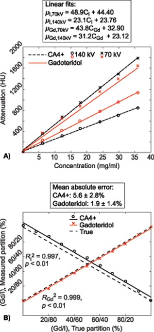
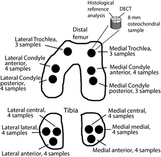
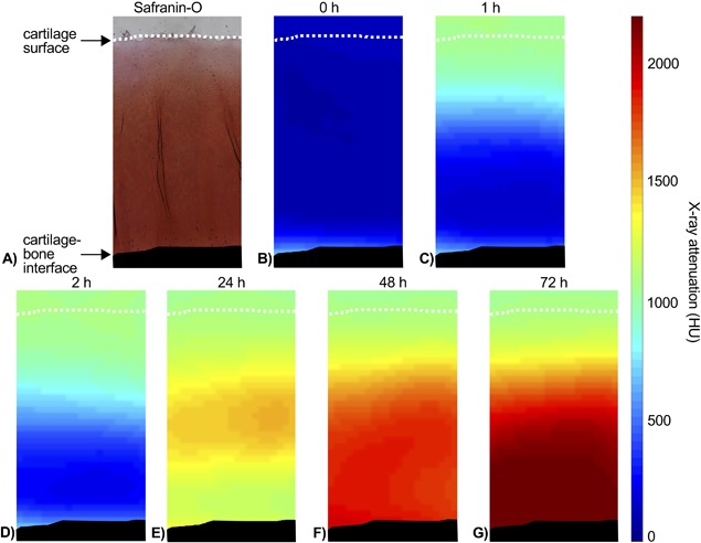
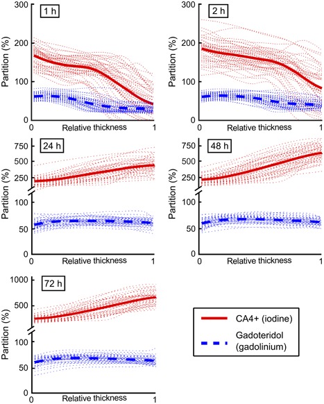
Assessment of cartilage composition via tomographic imaging is critical after cartilage injury to prevent post-traumatic osteoarthritis. Diffusion of cationic contrast agents in cartilage is affected by proteoglycan loss and elevated water content. These changes have opposite effects on diffusion and, thereby, reduce the diagnostic accuracy of cationic agents. Here, we apply, for the first time, a clinical full-body CT for dual contrast imaging of articular cartilage. We hypothesize that full-body CT can simultaneously determine the diffusion and partitioning of cationic and non-ionic contrast agents and that normalization of the cationic agent partition with that of the non-ionic agent minimizes the effect of water content and tissue permeability, especially at early diffusion time points. Cylindrical (d = 8 mm) human osteochondral samples (n = 45; four cadavers) of a variable degenerative state were immersed in a mixture of cationic iodinated CA4+ and non-charged gadoteridol contrast agents and imaged with a full-body CT scanner at various time points. Determination of contrast agents' distributions within cartilage was possible at all phases of diffusion. At early time points, gadoteridol, and CA4+ distributed throughout cartilage with lower concentrations in the deep cartilage. At ≥24 h, the gadoteridol concentration remained nearly constant, while the CA4+ concentration increased toward deep cartilage. Normalization of the CA4+ partition with that of gadoteridol significantly (p < 0.05) enhanced correlation with proteoglycan content and Mankin score at the early time points. To conclude, the dual contrast technique was found advantageous over single contrast imaging enabling more sensitive diagnosis of cartilage degeneration. © 2019 The Authors. Journal of Orthopaedic Research Published by Wiley Periodicals, Inc. J Orthop Res 9999:1-12, 2019.
Keywords: cartilage; cationic contrast agent; contrast enhanced computed tomography; dual contrast agent; dual energy computed tomography.
© 2019 The Authors. Journal of Orthopaedic Research Published by Wiley Periodicals, Inc.
Figures
References
-
- Buckwalter JA, Mankin HJ. 1997. Instructional Course Lectures, The American Academy of Orthopaedic Surgeons −Articular cartilage. Part II: degeneration and osteoarthrosis, repair, regeneration, and transplantation. J Bone Jt Surg − Am 79:612–632.
-
- Bay‐Jensen A‐C, Hoegh‐Madsen S, Dam E, et al. 2010. Which elements are involved in reversible and irreversible cartilage degradation in osteoarthritis? Rheumatol Int 30:435–442. - PubMed
Publication types
MeSH terms
Substances
LinkOut - more resources
Full Text Sources
