Review
. 2019 Apr;39(4):593-602.
doi: 10.1161/ATVBAHA.118.311906.

Brain Vasculature and Cognition

Affiliations
Review

Brain Vasculature and Cognition

Abdelrahman Y Fouda et al. Arterioscler Thromb Vasc Biol. 2019 Apr.

Abstract

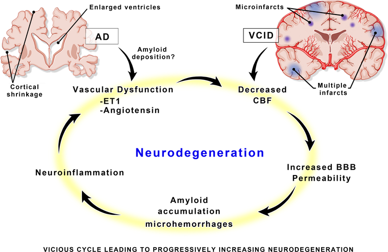
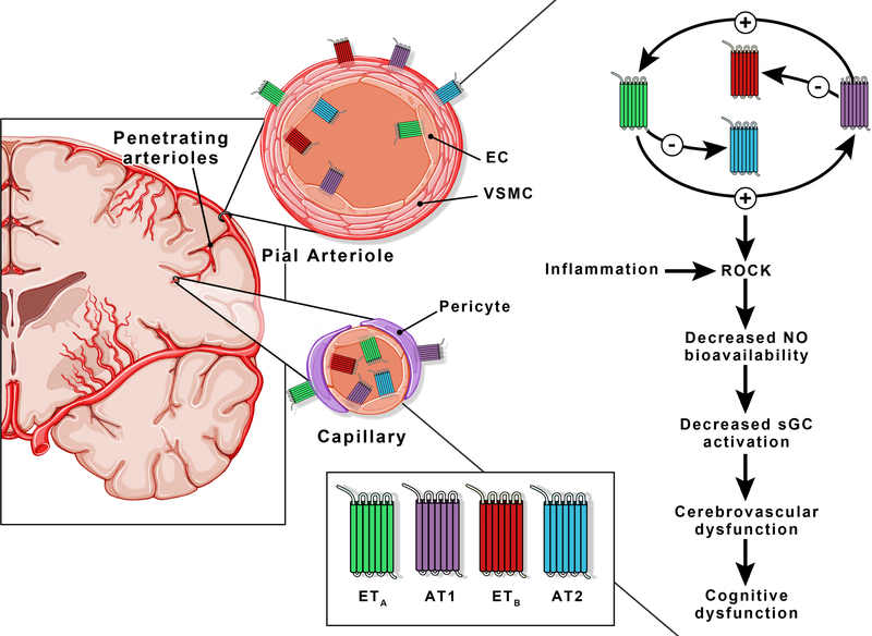
There is a complex interaction between the brain and the cerebral vasculature to meet the metabolic demands of the brain for proper function. Preservation of cerebrovascular function and integrity has a central role in this sophisticated communication within the brain, and any derangements can have deleterious acute and chronic consequences. In almost all forms of cognitive impairment, from mild to Alzheimer disease, there are changes in cerebrovascular function and structure leading to decreased cerebral blood flow, which may initiate or worsen cognitive impairment. In this focused review, we discuss the contribution of 2 major vasoactive pathways to cerebrovascular dysfunction and cognitive impairment in an effort to identify early intervention strategies.

Keywords: brain; cardiovascular system; cognitive dysfunction; endothelins; renin-angiotensin system.


Figures

Figure 1.
Figure 1.
A vicious cycle of pathologic neurovascular changes in the brain, leads to a malignantly progressive burden of neurodegeneration. Although Alzheimer’s disease (AD) and vascular cognitive impairment/dementia (VCID) may enter the cycle at different initiating points, the mediators of damage are similar and occur long before clinical symptoms present. BBB = blood–brain barrier, ET1 = endothelin 1, CBF = cerebral blood flow.
Figure 2.
Figure 2.
ET and Ang II receptor localization on the cerebrovascular networks. Both ET (ETA and ETB) and Ang II (AT1 and AT2) receptors are present on endothelial and smooth muscle cells of the pial vessels. A distinction at the capillary level is the co-presence of ETA and ETB receptors on endothelial cells. There is a bidirectional crosstalk between the ET system and RAS: ET-1 upregulates deleterious AT1 and downregulates protective AT2 receptor expression while Ang II upregulates deleterious ETA and downregulates protective ETB receptor expression. AT1 and ETA receptor signaling activates ROCK, decreases NO bioavailability and sGC signaling ultimately resulting in reduced CBF and cognitive deficits. EC = endothelial cells, VSMC = vascular smooth muscle cells, ROCK = Rho-associated protein kinase, NO = nitric oxide, sGC = Soluble guanylyl cyclase.

References

    1. Iadecola C The neurovascular unit coming of age: A journey through neurovascular coupling in health and disease. Neuron. 2017;96:17–42 - PMC - PubMed
    1. Sweeney MD, Zhao Z, Montagne A, Nelson AR, Zlokovic BV. Blood-brain barrier: From physiology to disease and back. Physiological reviews. 2019;99:21–78 - PMC - PubMed
    1. Sweeney MD, Kisler K, Montagne A, Toga AW, Zlokovic BV. The role of brain vasculature in neurodegenerative disorders. Nature neuroscience. 2018;21:1318–1331 - PMC - PubMed
    1. Sweeney MD, Sagare AP, Zlokovic BV. Blood-brain barrier breakdown in alzheimer disease and other neurodegenerative disorders. Nature reviews. Neurology 2018;14:133–150 - PMC - PubMed
    1. Gorelick PB, Furie KL, Iadecola C, et al. Defining optimal brain health in adults: A presidential advisory from the american heart association/american stroke association. Stroke. 2017;48:e284–e303 - PMC - PubMed

Publication types

MeSH terms

Substances