. 2019 Apr;39(4):e118-e129.
doi: 10.1161/ATVBAHA.119.312407.

Tuning the Thromboinflammatory Response to Venous Flow Interruption by the Ectonucleotidase CD39

Affiliations

Tuning the Thromboinflammatory Response to Venous Flow Interruption by the Ectonucleotidase CD39

Anuli C Anyanwu et al. Arterioscler Thromb Vasc Biol. 2019 Apr.

Abstract

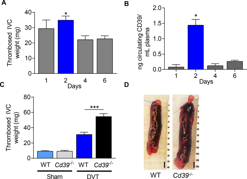
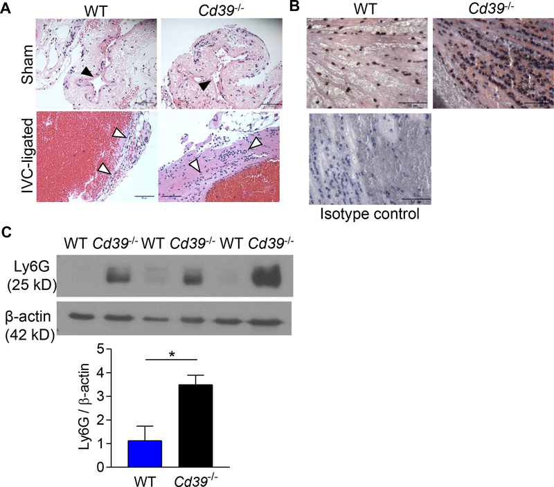
Objective- Leukocyte flux contributes to thrombus formation in deep veins under pathological conditions, but mechanisms that inhibit venous thrombosis are incompletely understood. Ectonucleotide di(tri)phosphohydrolase 1 ( ENTPD1 or Cd39), an ectoenzyme that catabolizes extracellular adenine nucleotides, is embedded on the surface of endothelial cells and leukocytes. We hypothesized that under venous stasis conditions, CD39 regulates inflammation at the vein:blood interface in a murine model of deep vein thrombosis. Approach and Results- CD39-null mice developed significantly larger venous thrombi under venous stasis, with more leukocyte recruitment compared with wild-type mice. Gene expression profiling of wild-type and Cd39-null mice revealed 76 differentially expressed inflammatory genes that were significantly upregulated in Cd39-deleted mice after venous thrombosis, and validation experiments confirmed high expression of several key inflammatory mediators. P-selectin, known to have proximal involvement in venous inflammatory and thrombotic events, was upregulated in Cd39-null mice. Inferior vena caval ligation resulted in thrombosis and a corresponding increase in both P-selectin and VWF (von Willebrand Factor) levels which were strikingly higher in mice lacking the Cd39 gene. These mice also manifest an increase in circulating platelet-leukocyte heteroaggregates suggesting heterotypic crosstalk between coagulation and inflammatory systems, which is amplified in the absence of CD39. Conclusions- These data suggest that CD39 mitigates the venous thromboinflammatory response to flow interruption.

Keywords: P-selectin; endothelial cells; inflammation; leukocytes; venous thrombosis.


Conflict of interest statement

DISCLOSURES

The authors declare no relevant disclosures.

Figures

Figure 1.
Figure 1.. Characterization of thrombus weight in WT and Cd39−/− mice 48 hr after stasis-induced thrombosis.
(A) Thrombosed IVC weight in WT mice 1–6 days following IVC ligation-induced venous thrombosis (1d, n=7; 2d, n=13; 4d, n=9; 6d, n=11, *P < 0.05 vs. 4d and 6d). (B) Circulating plasma soluble CD39 levels in WT mice following IVC-ligation (1d, n=3; 2d, n=3; 4d, n=3; 6d, n=6). (C) Thrombus-containing IVC weight in sham-operated mice and stasis-induced venous thrombosis in WT and Cd39−/− mice at 48 hours. Sham: WT, n=8; Cd39−/− n=8. IVC-ligated: WT, n=12; Cd39−/− n=7. (D) Representative photomicrographs of stasis-induced venous thrombosis of WT and Cd39−/− mice at 48 hours following IVC ligation. Scale bar, 1 mm. Data are shown as mean ± SEM. *P<0.05, **P<0.01, ***P<0.005.
Figure 2.
Figure 2.. Increased leukocyte recruitment to the thrombosed IVC in WT and Cd39−/− mice.
(A) Hematoxylin and eosin stained inferior vena cava section of 48 hour sham operated- and thrombosed-IVC of WT and Cd39−/− mice. Black arrowheads=IVC; white arrowheads=recruited leukocytes. Scale bar, 50 μm; representative sections, n=5 each. (B) Neutrophil staining (Ly6G, brown) in thrombosed veins of WT and Cd39−/− mice counterstained with hematoxylin. Scale bar, 50 μm; representative sections, n=3 each. (C) Immunoblot for Ly6G in venous thrombi from WT and Cd39−/− mice. N=3, each; *P<0.05
Figure 3.
Figure 3.. Gene expression patterns demonstrate distinct vessel wall thrombo-inflammatory signaling pathways in WT and Cd39−/− mice in venous thrombosis.
Inferior vena cava gene expression patterns due to CD39 deficiency or stasis-induced thrombosis. Venn diagrams illustrating the number of differentially expressed genes (probe sets; fold change ≥2; P≤0.001) in each pairwise comparison of IVCs between (A) WT sham vs. WT IVC-ligated; and (B) WT IVC-ligated vs. Cd39−/− IVC-ligated mice (n=4 pooled IVCs, each). Hierarchical clustering of a subset of dynamically expressed genes involved in inflammation and immunity following venous thrombosis in (C) WT mice; and (D) following Cd39−/− deficiency. (E) Network analysis of co-expressed key inflammatory genes under venous stasis thrombosis conditions in Cd39−/− compared with WT mice. Red: upregulation; green: downregulation.
Figure 4.
Figure 4.. Validation of microarray results reveals quantitative RT-PCR analysis of WT and Cd39−/− mice 48 hr following IVC ligation.
Gene expression of pro-inflammatory mediators and chemokines in vena cava of WT and Cd39−/− mice 48 hours after IVC ligation using qRT-PCR normalized to β-actin expression. Data shown as mean ± SEM, n≥4, each. *P<0.05, **P<0.01, ***P<0.001.
Figure 5.
Figure 5.. CD39-deficiency increases local and circulating P-selectin and von Willebrand Factor expression.
Plasma soluble (A) P-selectin and (B) circulating VWF levels in WT and Cd39−/− mice. *P<0.05, ***P<0.001; n=3–6 (A) and n=11–31 (B). Localization of vein wall and thrombus (C) P-selectin and (D) VWF by immunohistochemistry in sham and IVC-ligated WT and Cd39−/− mice. Black arrowheads, P-selectin; white arrowheads, VWF; scale bar, 50 μm.
Figure 6.
Figure 6.. Increased circulating activated platelet-leukocyte aggregates in Cd39−/− mice following venous thrombosis.
Representative flow cytometric analysis of myeloid-derived leukocytes in whole blood from WT and Cd39−/− sham-operated or IVC-ligated mice. (A) activated CD62+ platelet-CD11b+ leukocyte aggregates in Cd39−/− mice (76.3% ± 12.3; n=3) and WT mice (52.6% ± 10.2; n=4. P<0.001), and (B) activated CD62P+ platelet-Gr-1+ leukocyte aggregates. Quantitative cell count analysis of (C) CD11b+/CD41+/CD62P+ cells, and (D) GR-1+/CD41+/CD62P+ cells in WT and Cd39−/− sham operated or IVC-ligated mice 48 hours post IVC-ligation. Data are shown are n=3–5, mean ± SD. *P<0.05, ***P<0.0001.

Similar articles

Cited by

References

    1. Silverstein MD, Heit JA, Mohr DN, Petterson TM, O’Fallon WM, Melton LJ 3rd. Trends in the incidence of deep vein thrombosis and pulmonary embolism: A 25-year population-based study. Arch Intern Med. 1998;158:585–593 - PubMed
    1. Bureau. USC. Monthly populations estimates for the united states: April 1, 2010 to december 1, 2012. Population Estimates. 2012;2012
    1. Yusuf HR, Tsai J, Atrash HK, Boulet S, Gross SD. Venous thromboembolism in adult hospitalizations-united states, 2007–2009. Atlanta, Ga: U.S. Dept. of Health and Human Services, Centers for Disease Control and Prevention; 2012.
    1. Center for Diseases Control. Venous thromboembolism.2018
    1. Ocak G, Vossen CY, Verduijn M, Dekker FW, Rosendaal FR, Cannegieter SC, Lijfering WM. Risk of venous thrombosis in patients with major illnesses: Results from the mega study. J Thromb Haemost. 2012 - PubMed

Publication types

MeSH terms