. 2019 May;33(5):6011-6022.
doi: 10.1096/fj.201801993R. Epub 2019 Feb 28.

Salivary gland immunization via Wharton's duct activates differential T-cell responses within the salivary gland immune system

Affiliations

Salivary gland immunization via Wharton's duct activates differential T-cell responses within the salivary gland immune system

Guangliang Liu et al. FASEB J. 2019 May.

Abstract

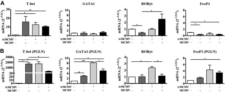
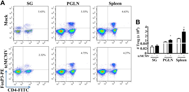
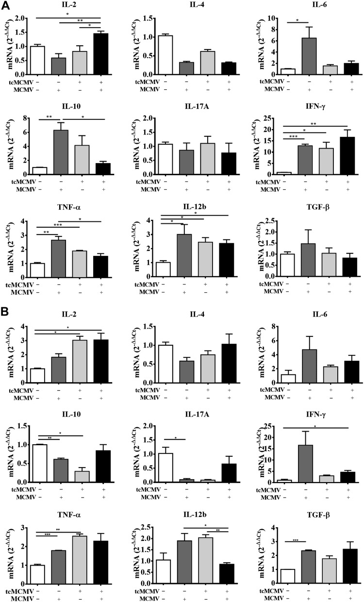
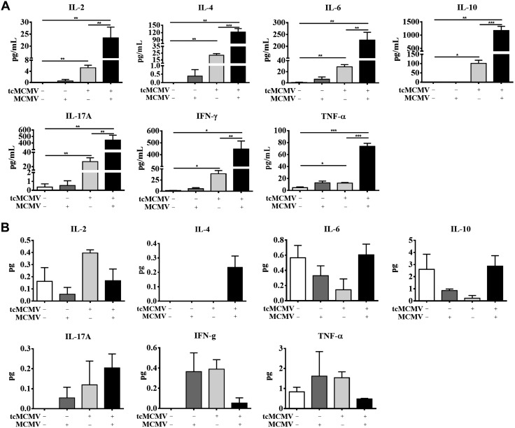
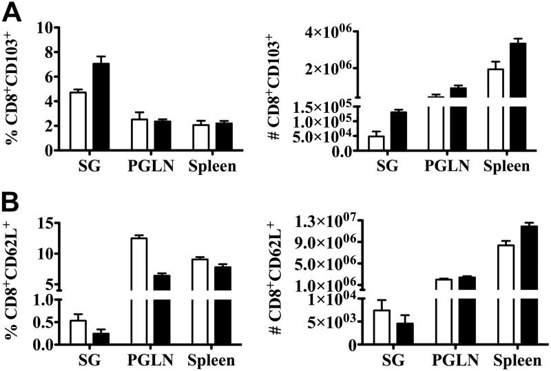
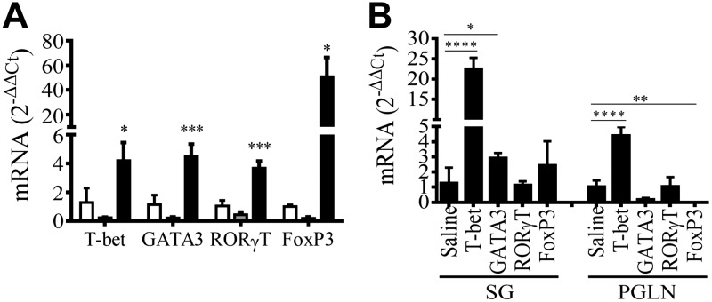
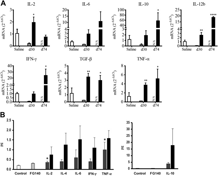
Salivary glands are a major component of the mucosal immune system that confer adaptive immunity to mucosal pathogens. As previously demonstrated, immunization of the submandibular gland with tissue culture-derived murine cytomegalovirus (tcMCMV) or replication-deficient adenoviruses expressing individual murine cytomegalovirus (MCMV) genes protected mice against a lethal MCMV challenge. Here, we report that salivary gland inoculation of BALB/cByJ mice with tcMCMV or recombinant adenoviruses differentially activates T helper (Th)1, -2, and -17 cells in the salivary glands vs. the associated lymph nodes. After inoculation with tcMCMV, lymphocytes from the submandibular gland preferentially express the transcription factor T-cell-specific T-box transcription factor (T-bet), which controls the expression of the hallmark Th1 cytokine, IFN-γ. Lymphocytes from the periglandular lymph nodes (PGLNs) express both T-bet and GATA-binding protein 3 (GATA3), which promotes the secretion of IL-4, -5, and -10 from Th2 cells. In contrast, after inoculation with replication-deficient adenoviruses, lymphocytes from the submandibular gland express T-bet, GATA3, and RAR-related orphan receptor γ, thymus-specific isoform (RORγt) (required for differentiation of Th17 cells) and forkhead box P3 (Foxp3) (required for the differentiation of regulatory T cells). Lymphocytes from the PGLNs were not activated. The differential induction of Th responses in the salivary gland vs. the PGLNs after inoculation with attenuated virus vs. a nominal protein antigen supports the use of the salivary as an alternative mucosal route for administering vaccines.-Liu, G., Zhang, F., Wang, R., London, S. D., London, L. Salivary gland immunization via Wharton's duct activates differential T-cell responses within the salivary gland immune system.

Keywords: T helper cells; mucosal immunity; vaccine development.


Conflict of interest statement

The authors thank Dr. John D. Shanley and Dr. Carol A. Wu (University of Connecticut, Farmington, CT, USA) for providing recombinant adenovirus vectors and cell lines. This project was supported by U.S. National Institutes of Health/National Institute of Dental and Craniofacial Research Grant 1R01 DE016652 (to S.D.L.) and 1R15DE024313 (to S.D.L.). The authors declare no conflicts of interest.

Figures

Figure 1
Figure 1
Retrograde perfusion of the submandibular salivary gland with tcMCMV resulted in the expression of transcription factors associated with Th1/Th2/Th17 cytokines after a lethal challenge with MCMV. BALB/cByJ mice were inoculated with saline (mock) or 105 PFU/mouse tcMCMV via retrograde perfusion of the salivary gland. On d 28 after inoculation, saline mice were challenged intraperitoneally with 2 × 10⁴ PFU/mouse MCMV, and tcMCMV mice were challenged intraperitoneally with 5 × 10⁴ PFU/mouse MCMV. RNA was prepared from the PGLNs and the lymphocytes obtained from the whole salivary glands after removal of the associated lymph nodes in saline control (mock) (−/−), primed (+/−), or postchallenge (−/+ or +/+) mice on d 14 and analyzed for expression of the indicated transcription factors by qRT-PCR. A) Salivary glands. B) PGLNs. Data are the means ± sd of 5 mice/group. *P < 0.05, **P < 0.01, ***P < 0.001.
Figure 2
Figure 2
Retrograde perfusion of the submandibular gland with tcMCMV modulated CD4+Foxp3+ Treg cells. BALB/cByJ mice were inoculated with either saline (mock) or 105 PFU/mouse tcMCMV via retrograde perfusion of the salivary gland. Single-cell suspensions were prepared from lymphocytes obtained from the whole salivary glands, PGLNs, and spleens on d 14 after inoculation and analyzed for the coexpression of the T-cell surface marker CD4 and the intracellular marker Foxp3. A) Dot plots demonstrate lymphocytes stained with CD4 (x axis) and Foxp3 (y axis). The percentage of dual-staining cells is shown in the upper right quadrant. B) The histogram demonstrates the absolute numbers of CD4+Foxp3+ Treg cells from saline- (open bars) and tcMCMV- (solid bar) inoculated mice. Data are the means ± sd from 5 individual mice/group. *P < 0.05 vs. saline
Figure 3
Figure 3
Retrograde perfusion of the submandibular gland with tcMCMV resulted in Th1/Th2/Th17 cytokine mRNA expression after a lethal challenge with MCMV. BALB/cByJ mice were inoculated with saline (mock) or 105 PFU/mouse tcMCMV via retrograde perfusion of the salivary gland. On d 28 after inoculation, saline mice were challenged intraperitoneally with 2 × 10⁴ PFU/mouse MCMV and tcMCMV mice were challenged intraperitoneally with 5 × 10⁴ PFU/mouse MCMV. RNA was prepared from PGLNs and lymphocytes obtained from whole salivary glands after removal of the associated lymph nodes in saline control (mock) (−/−), primed (+/−), or postchallenge (−/+ or +/+) mice on d 14 and analyzed for expression of the indicated cytokines/chemokines by qRT-PCR. A) Salivary glands. B) PGLNs. Data are the means ± sd of 5 mice/group. *P < 0.05, **P < 0.01, ***P < 0.001.
Figure 4
Figure 4
Retrograde perfusion of the submandibular gland with tcMCMV resulted in Th1/Th2/Th17 cytokine protein expression after a lethal challenge with MCMV. BALB/cByJ mice were inoculated with saline (mock) or 105 PFU/mouse tcMCMV via retrograde perfusion of the salivary gland. On d 28 after inoculation, saline mice were challenged intraperitoneally with 2 × 10⁴ PFU/mouse MCMV and tcMCMV mice were challenged with 5 × 10⁴ PFU/mouse MCMV. Serum and saliva samples were collected from saline control (mock) (−/−), primed (+/−), or postchallenge (−/+ or +/+) mice on d 14 and subjected to a Th1/Th2/Th17 cytokine bead array assay. A) Serum (pg/ml). B) Saliva (total pg) recovered to normalize for differences in the volume of saliva collected from individual animals. Data are the means ± sd of 5 mice/group. *P < 0.05, **P < 0.01, ***P < 0.001.
Figure 5
Figure 5
Retrograde perfusion of the submandibular gland with tcMCMV resulted in increased expression of integrin αE. BALB/cByJ mice were inoculated with saline (mock) or 105 PFU/mouse tcMCMV via retrograde perfusion of the salivary gland. On d 28 after inoculation, saline mice were challenged intraperitoneally with 2 × 10⁴ PFU/mouse MCMV and tcMCMV mice were challenged intraperitoneally with 5 × 10⁴ PFU/mouse MCMV. Single-cell suspensions of lymphocytes were prepared from the whole salivary glands, PGLNs, and spleens on d 14 postchallenge and analyzed for the coexpression of the T-cell surface marker CD8 and the adhesion molecules CD62L or -103. A) The histogram demonstrates the percentage and absolute numbers of CD8+CD103+ in mock (−/−) (open bars) or tcMCMV postchallenge with MCMV (solid bar). B) The histogram demonstrates the absolute numbers of CD8+CD62L+ in mock (−/−) (open bars) or tcMCMV postchallenge with MCMV (solid bar). Data are the mean ± sd from 5 individual mice/group.
Figure 6
Figure 6
Retrograde perfusion of the submandibular salivary gland with replication-deficient recombinant adenovirus expressing MCMV genes resulted in the expression of transcription factors associated with Th1/Th2/Th17 responses. BALB/cByJ mice were inoculated (d 0) and boosted on d 30 with either saline, 106 PFU/mouse of FG140, or a combination of Ad-gpB, -gpH, and -IE1 (106 PFU/mouse of each recombinant virus, Ad-Combo) via retrograde perfusion of the submandibular salivary gland. On d 30 after boost, mice inoculated with saline were challenged intraperitoneally with saline and mice inoculated with FG140 or Ad-Combo were challenged intraperitoneally with 5 × 10⁴ PFU/mouse MCMV. RNA was prepared from the whole salivary glands after removal of the associated lymph nodes and from the PGLNs and were analyzed for expression of the indicated transcription factors by qRT-PCR. A) Salivary gland RNA was analyzed from mice inoculated (d 0) and boosted on d 30 (prechallenge) with saline (mock, open bars), 106 PFU/mouse of FG140 (negative control, gray bars), or with Ad-Combo (solid bars) for expression of Th-related transcription factors. B) RNA from the salivary glands and PGLNs at 2 wk postchallenge was analyzed from mice inoculated and boosted (d 0, d 30) with Ad-Combo and challenged with 5 × 10⁴ PFU/mouse MCMV (d 72) (solid bars) for expression of Th-related transcription factors. Data are the means ± sd of ≤5 mice/group. *P < 0.05, **P < 0.01, ***P < 0.001, ****P < 0.0001.
Figure 7
Figure 7
Retrograde perfusion of the submandibular salivary gland with replication-deficient recombinant adenovirus expressing MCMV genes resulted in the expression of cytokines associated with Th1/Th2/Th17 responses. BALB/cByJ mice were inoculated (d 0) and boosted on d 30 with either saline, 106 PFU/mouse of FG140, or a combination of Ad-gpB, -gpH, and -IE1 (106 PFU/mouse of each recombinant virus, Ad-Combo) via retrograde perfusion of the submandibular salivary gland. On d 30 after boost, mice inoculated with saline were challenged intraperitoneally with saline, and mice inoculated with FG140 or Ad-Combo were challenged intraperitoneally with 5 × 10⁴ PFU/mouse MCMV. A) RNA was prepared from the whole salivary glands after removal of the associated lymph nodes and was analyzed for Th-related cytokine expression at d 30 after primary inoculation and boost (prechallenge) and at 2 wk post-boost/challenge (d 74). Saline (open bars), FG140 (negative control, gray bars), and Ad-Combo (solid bars). Data were not available for FG140-inoculated mice after lethal challenge (d 74) due to death. B) Saliva samples were collected from mice inoculated with saline (control, open bars), from FG140 (negative control, gray bars), from Ad-Combo at d 30 after primary inoculation and boost (prechallenge, hashed bars), and from Ad-Combo mice challenged intraperitoneally with MCMV (d 74 postchallenge, solid bars) and were analyzed via a Th1/Th2/Th17 cytokine bead array assay. Saliva was expressed as total picograms recovered to normalize for differences in the volume of saliva collected from individual animals. Data are the mean ± sd of ≤5 mice/group. *P < 0.05, **P < 0.01, ****P < 0.0001.

Similar articles

Cited by

References

    1. Takahashi I., Nochi T., Yuki Y., Kiyono H. (2009) New horizon of mucosal immunity and vaccines. Curr. Opin. Immunol. 21, 352–358 - PubMed
    1. Brandtzaeg P. (2007) Induction of secretory immunity and memory at mucosal surfaces. Vaccine 25, 5467–5484 - PubMed
    1. Neutra M. R., Kozlowski P. A. (2006) Mucosal vaccines: the promise and the challenge. Nat. Rev. Immunol. 6, 148–158 - PubMed
    1. Tucker S. N., Lin K., Stevens S., Scollay R., Bennett M. J., Olson D. C. (2004) Salivary gland genetic vaccination: a scalable technology for promoting distal mucosal immunity and heightened systemic immune responses. Vaccine 22, 2500–2504 - PubMed
    1. Grewal J. S., Pilgrim M. J., Grewal S., Kasman L., Werner P., Bruorton M. E., London S. D., London L. (2011) Salivary glands act as mucosal inductive sites via the formation of ectopic germinal centers after site-restricted MCMV infection. FASEB J. 25, 1680–1696 - PMC - PubMed

Publication types