. 2019 May;33(5):6390-6401.
doi: 10.1096/fj.201802270R. Epub 2019 Feb 28.

Naturally presented HLA class I-restricted epitopes from the neurotrophic factor S100-β are targets of the autoimmune response in type 1 diabetes

Affiliations

Naturally presented HLA class I-restricted epitopes from the neurotrophic factor S100-β are targets of the autoimmune response in type 1 diabetes

Cristina Calviño-Sampedro et al. FASEB J. 2019 May.

Abstract

Type 1 diabetes (T1D) results from the destruction of pancreatic β-cells by the immune system, and CD8+ T lymphocytes are critical actors in this autoimmune response. Pancreatic islets are surrounded by a mesh of nervous cells, the peri-insular Schwann cells, which are also targeted by autoreactive T lymphocytes and express specific antigens, such as the neurotrophic factor S100-β. Previous work has shown increased proliferative responses to whole S100-β in both human T1D patients and the nonobese diabetic (NOD) mouse model. We describe for the first time naturally processed and presented epitopes (NPPEs) presented by class I human leukocyte antigen-A*02:01 (A2.1) molecules derived from S100-β. These NPPEs triggered IFN-γ responses more frequently in both newly diagnosed and long-term T1D patients compared with healthy donors. Furthermore, the same NPPEs are recognized during the autoimmune response leading to diabetes in A2.1-transgenic NOD mice as early as 4 wk of age. Interestingly, when these NPPEs are used to prevent diabetes in this animal model, an acceleration of the disease is observed together with an exacerbation in insulitis and an increase in S100-β-specific cytotoxicity in vaccinated animals. Whether these can be used in diabetes prevention needs to be carefully evaluated in animal models before use in future clinical assays.-Calviño-Sampedro, C., Gomez-Tourino, I., Cordero, O. J., Reche, P. A., Gómez-Perosanz, M., Sánchez-Trincado, J. L., Rodríguez, M. Á., Sueiro, A. M., Viñuela, J. E., Calviño, R. V. Naturally presented HLA class I-restricted epitopes from the neurotrophic factor S100-β are targets of the autoimmune response in type 1 diabetes.

Keywords: S100β peptide epitopes; autoantigen; cytotoxic lymphocytes; immunotherapy; peri-insular Schwann cells.


Conflict of interest statement

The authors thank Dr. Sefina Arif (King’s College London, London, United Kingdom) for critically reviewing the manuscript. This work was funded by the Ministerio de Economía y Competitividad (Grant BIO2014-53091-C3-3-R to R.V.C.). During this work, I.G.-T. was supported by a Maria Barbeito predoctoral fellowship (Xunta de Galicia, La Coruña, Spain). During this work, C.C.-S. was supported by a Deputación da Coruña grant (2012–2013 and 2016–2017). The authors declare no conflicts of interest.

Figures

Figure 1
Figure 1
MS analysis of in vitro proteasome digestion of purified S100-β. A, B) Spectra corresponding to m/z range of 917–1564 (A) and 1556–3145 (B) are shown. Data for S100-β incubated with no proteasome (top panels), inactive proteasome (middle panels), or active proteasome (bottom panels) are shown. Unique m/z present only in samples of S100-β incubated with active proteasome were subsequently identified by tandem MS and analyzed by Mascot (arrows). C) Sequences of the 9 most intense fragments generated by in vitro digestion of S100-β with purified 20S proteasome are aligned with the amino acid sequence of the antigen. The m/z for each fragment is shown on the left. The carboxy-terminal amino acid is underlined. The sequences of potential peptide epitopes eluted from A2.1 are shown at the bottom (carboxy-terminal amino acid is underlined only if the fragment has been generated by the proteasome and identified by MS). The carboxy-terminal ends of peptides S10010–18 (ALIDVFHQY) and S10020–28 (GREGDKHKL) are generated by the proteasome, making them potential class I–restricted peptide epitopes.
Figure 2
Figure 2
IFN-γ ELISPOT analysis of responses against S10010–18 and S10020–28 peptide epitopes. A) ROC curves were performed to discriminate optimal cutoff values between responders and nonresponders, giving an SI cutoff of 1.85 and 1.75 for S10010–18 and S10020–28, respectively. An SI ≥2 was used as a cutoff. B) A representative response from a T1D patient is shown (in parentheses, spot mean number ± sd of triplicate wells) for S10010–18 and S10020–28. A peptide mixture containing known peptide epitopes derived from viral proteins (CEF) was included as a positive control. Spontaneous responses were determined by PBMC culture in medium alone (culture medium) or medium containing 0.5% DMSO. C–F) IFN-γ secretion in response to S10010–18 (C, E) and S10020–28 (D, F) by PBMCs from HDs, all T1D patients (T1D), ND T1D patients (T1D-ND), and LS T1D patients (T1D-LS). Black symbols: HLA-A2.1–negative subjects; red symbols: HLA-A2.1–positive subjects. Kruskal-Wallis test with Dunn’s post hoc test (C, D). *P < 0.05, **P < 0.01, ***P < 0.001. G) Responses against both S10010–18 and S10020–28 were more frequently detected among T1D patients (ND T1D: 57.1%; LS T1D: 60%) compared with HDs (11.1%). Two-tailed Fisher’s exact test, P = 0.0045. There is also a positive correlation between the responses to both peptides (r = 0.66, Spearman’s correlation, P < 0.0001). The dashed line (CG) represents the threshold.
Figure 3
Figure 3
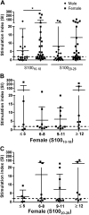
Spontaneous autoimmune responses against S10010-18 and S10020-28 in A2.1-transgenic NOD mice. A) Spontaneous autoimmune responses against S10010–18 and S10020–28 were determined in splenocytes by IFN-γ ELISPOT in male (n = 22; 10.32 ± 4.76 wk of age, mean ± sd) and female (n = 31; 9.83 ± 3.97, mean ± sd) A2.1-transgenic NOD mice. Data shown are the SIs (y axis) calculated as the ratio between the number of spots with peptide and the number of spots in culture medium only. SIs ≥2 were considered positive responses. Median responses were higher in females compared with males for S10010–18. B, C) Mann-Whitney U test. P = 0.035. Spontaneous responses against S10010–18 (B) and S10020–28 (C) according to age were determined in female mice (n = 31; 5–10 animals/age group). Positive responses (SI ≥2) can be detected as early as 5 wk of age for both peptide epitopes. The dashed line represents the threshold.
Figure 4
Figure 4
T1D development in A2.1-transgenic NOD mice vaccinated intraperitoneally with S10010–18 and S10020–28. A) Animals were vaccinated with 100 μg each of S10010–18 and S10020–28 per dose (n = 10; continuous line) starting at 4 wk of age and then every 2–3 wk until they developed diabetes or reached 30 wk of age. Animals inoculated with saline (n = 12; dotted line) were used as controls. Vaccination with S10010–18 and S10020–28 does not seem to significantly modify either the incidence or the kinetics of diabetes development in A2.1-transgenic NOD mice. Log-rank, Mantel-Cox, P = 0.29. B) Insulitis in A2.1-transgenic NOD mice vaccinated with S100-β–derived NPPEs or inoculated with saline. The percentage of islets heavily infiltrated (scores 3 and 4) is significantly higher in S100-β–vaccinated animals compared with those inoculated with saline. Fisher’s exact test; P = 0.0054. C, D) IFN-γ secretion in response to S10010–18 (C) and S10020–28 (D) in A2.1-transgenic NOD mice vaccinated with the S100-β–derived NPPEs (S100-β), saline (saline), or nonmanipulated (spontaneous). In all cases, splenocytes were harvested at disease diagnosis or at 30 wk of age. Neither frequency of positive responses (Fisher’s exact test, P > 0.05) nor median SI responses (Kruskal-Wallis test, P = 0.4) were significantly different in S100-β–immunized mice compared with spontaneous and saline-inoculated mice.
Figure 5
Figure 5
T1D diabetes development in A2.1-transgenic NOD mice vaccinated intraperitoneally with low doses of S10010–18 and S10020–28. A) Animals were vaccinated with 10 μg each of S10010–18 and S10020–28 per dose (n = 10; continuous line) starting at 4 wk of age and then every 2–3 wk until they developed diabetes or reached 30 wk of age. Animals inoculated with saline (n = 12; black dotted line) were used as controls. Spontaneous disease development in nonmanipulated females (n = 34; gray dotted line) is also shown. Vaccination with S10010-18 and S10020-28 significantly accelerates the kinetics of diabetes development in A2.1-transgenic mice compared with both saline-treated (log-rank, Mantel-Cox, P = 0.048) and nonmanipulated (log-rank, Mantel-Cox, P = 0.02) female mice. B) Insulitis in A2.1-transgenic NOD mice vaccinated with S100-β–derived NPPEs or inoculated with saline. The percentage of islets heavily infiltrated (scores 3 and 4) is significantly higher in S100-β–vaccinated animals compared with those inoculated with saline. Fisher’s exact test; P = 0.011. C, D) IFN-γ secretion in response to S10010–18 (C) and S10020–28 (D) in A2.1-transgenic NOD mice vaccinated with the S100-β–derived NPPEs (S100-β), saline (saline), or nonmanipulated (spontaneous). In all cases, splenocytes were harvested at disease diagnosis or at 30 wk of age. Neither frequency of positive responses (Fisher’s exact test, P > 0.05) nor median SI responses (P = 0.24 for S10010–18 and P = 0.097 for S10020–28; Kruskal-Wallis test) were significantly different in S100-β–immunized mice compared with spontaneous and saline-inoculated mice. E) Splenocytes from animals vaccinated with S100-β–derived NNPEs (black line) show a higher specific cytotoxicity against targets incubated with both S100-β–derived NPPEs compared with that seen in splenocytes from animals inoculated with saline (dotted line). Mann-Whitney U test, P < 0.05 for all effector:target (E:T) ratios.

References

    1. Taplin C. E., Barker J. M. (2008) Autoantibodies in type 1 diabetes. Autoimmunity 41, 11–18 - PubMed
    1. Coppieters K. T., Dotta F., Amirian N., Campbell P. D., Kay T. W., Atkinson M. A., Roep B. O., von Herrath M. G. (2012) Demonstration of islet-autoreactive CD8 T cells in insulitic lesions from recent onset and long-term type 1 diabetes patients. J. Exp. Med. 209, 51–60 - PMC - PubMed
    1. Roep B. O., Peakman M. (2011) Diabetogenic T lymphocytes in human Type 1 diabetes. Curr. Opin. Immunol. 23, 746–753 - PubMed
    1. Anderson M. S., Bluestone J. A. (2005) The NOD mouse: a model of immune dysregulation. Annu. Rev. Immunol. 23, 447–485 - PubMed
    1. Liblau R. S., Wong F. S., Mars L. T., Santamaria P. (2002) Autoreactive CD8 T cells in organ-specific autoimmunity: emerging targets for therapeutic intervention. Immunity 17, 1–6 - PubMed

Publication types

MeSH terms