

An Examination of the Frequency of Paravascular Defects and Epiretinal Membranes in Eyes With Early Glaucoma Using En-face Slab OCT Images
- PMID: 30817498
- PMCID: PMC6400318
- DOI: 10.1097/IJG.0000000000001188
An Examination of the Frequency of Paravascular Defects and Epiretinal Membranes in Eyes With Early Glaucoma Using En-face Slab OCT Images
Abstract
Purpose: To examine the frequency of paravascular defects (PDs) and macular epiretinal membranes (ERMs) in eyes categorized as having mild glaucoma or glaucoma suspect using en-face slab analysis of optical coherence tomography (OCT) scans.
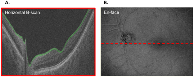
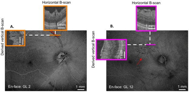
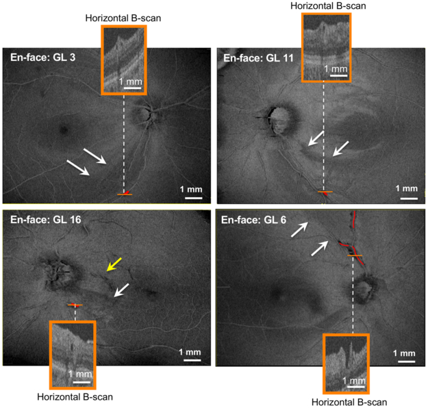
Materials and methods: Fifty-seven glaucomatous eyes, 44 low-risk suspect eyes, and 101 healthy control eyes were included in the study. The 101 glaucomatous and suspect eyes had a mean deviation better than -6 dB on the 24-2 visual field, and a spherical refractive error between±6 D or axial length <26.5 mm. Two OCT-graders masked to eye classification identified ERMs and PDs on en-face slab images of the macula and peripapillary retina using horizontal B-scans and derived vertical B-scans.
Results: Glaucomatous eyes had a significantly higher number of PDs and ERMs than healthy controls (PD, P<0.001; ERM, P=0.046) and low-risk glaucoma suspects (PD, P=0.004; ERM, P=0.043). PDs and/or ERMs were present in 16 of 57 (28.1%) glaucomatous eyes, 2 of 44 (4.5%) suspect eyes, and 3 of 101 (3.0%) control eyes. Further, PDs were present in 11 of the 57 (19.3%) glaucomatous eyes, 1 of the 44 (2.3%) suspect eyes and 0 of the 101 (0%) control eyes, ERMs were seen in 7 of the 57 (12.3%) glaucomatous eyes, 1 of the 44 (2.3%) suspects, and 3 of the 101 (3.0%) control eyes.
Conclusions: Eyes with early glaucoma have a higher frequency of PDs and ERMs than suspects or controls and exhibit PDs even in the absence of ERMs or high myopia.
Figures
Similar articles
-
Optical Coherence Tomography Can Be Used to Assess Glaucomatous Optic Nerve Damage in Most Eyes With High Myopia.J Glaucoma. 2020 Oct;29(10):833-845. doi: 10.1097/IJG.0000000000001631. J Glaucoma. 2020. PMID: 33006872 Free PMC article.
-
Defects Along Blood Vessels in Glaucoma Suspects and Patients.Invest Ophthalmol Vis Sci. 2016 Apr;57(4):1680-6. doi: 10.1167/iovs.15-18499. Invest Ophthalmol Vis Sci. 2016. PMID: 27054521 Free PMC article.
-
Inter-eye Asymmetry of Optical Coherence Tomography Angiography Vessel Density in Bilateral Glaucoma, Glaucoma Suspect, and Healthy Eyes.Am J Ophthalmol. 2018 Jun;190:69-77. doi: 10.1016/j.ajo.2018.03.026. Epub 2018 Mar 24. Am J Ophthalmol. 2018. PMID: 29580976 Free PMC article.
-
Progressive Macula Vessel Density Loss in Primary Open-Angle Glaucoma: A Longitudinal Study.Am J Ophthalmol. 2017 Oct;182:107-117. doi: 10.1016/j.ajo.2017.07.011. Epub 2017 Jul 20. Am J Ophthalmol. 2017. PMID: 28734815 Free PMC article.
-
The Association Between Clinical Features Seen on Fundus Photographs and Glaucomatous Damage Detected on Visual Fields and Optical Coherence Tomography Scans.J Glaucoma. 2017 May;26(5):498-504. doi: 10.1097/IJG.0000000000000640. J Glaucoma. 2017. PMID: 28333890 Free PMC article.
Cited by
-
A Simple Subjective Evaluation of Enface OCT Reflectance Images Distinguishes Glaucoma From Healthy Eyes.Transl Vis Sci Technol. 2021 May 3;10(6):31. doi: 10.1167/tvst.10.6.31. Transl Vis Sci Technol. 2021. PMID: 34036303 Free PMC article.
-
Long-Term Intraocular Pressure Fluctuation and Epiretinal Membrane in Patients with Glaucoma or Glaucoma Suspect.J Clin Med. 2024 Feb 17;13(4):1138. doi: 10.3390/jcm13041138. J Clin Med. 2024. PMID: 38398451 Free PMC article.
-
Optical Coherence Tomography Can Be Used to Assess Glaucomatous Optic Nerve Damage in Most Eyes With High Myopia.J Glaucoma. 2020 Oct;29(10):833-845. doi: 10.1097/IJG.0000000000001631. J Glaucoma. 2020. PMID: 33006872 Free PMC article.
-
Location and Extent of Paravascular Nerve Fiber Layer Clefts in Eyes with Epiretinal Membranes.J Clin Med. 2024 Sep 26;13(19):5731. doi: 10.3390/jcm13195731. J Clin Med. 2024. PMID: 39407790 Free PMC article.
-
Current Choroidal Imaging Findings in Central Serous Chorioretinopathy.Vision (Basel). 2020 Oct 16;4(4):44. doi: 10.3390/vision4040044. Vision (Basel). 2020. PMID: 33081096 Free PMC article. Review.
References
-
- Liu H-Y, Hsieh Y-T, Yang C-M. Paravascular abnormalities in eyes with idiopathic epiretinal membrane. Graefe’s Archive for Clinical and Experimental Ophthalmology. 2016;254:1723–1729. - PubMed
-
- Chihara E, Chihara K. Apparent cleavage of the retinal nerve fiber layer in asymptomatic eyes with high myopia. Graefe’s Archive for Clinical and Experimental Ophthalmology. 1992;230:416–420. - PubMed
-
- Komeima K, Kikuchi M, Ito Y, Terasaki H, Miyake Y. Paravascular inner retinal cleavage in a highly myopic eye. Arch Ophthalmol. 2005;123:1449–1450. - PubMed
-
- Hwang YH, Kim YF, KIm HK, Sohn YH. Characteristics of eyes with inner retinal cleavage. Graefes Arch Clin Exp Ophthalmol. 2015;253(2):215–220. - PubMed
-
- Muraoka Y, Tsujikawa A, Hata M, et al. Paravascular inner retinal defect associated with high myopia or epiretinal membrane. JAMA Ophthalmology. 2015;133:413–420. - PubMed
Publication types
MeSH terms
Grants and funding
LinkOut - more resources
Full Text Sources
Research Materials
Miscellaneous
