Review
. 2019 Feb 28;60(3):M31-M88.
doi: 10.1167/iovs.18-25967.

IMI - Report on Experimental Models of Emmetropization and Myopia

Affiliations
Review

IMI - Report on Experimental Models of Emmetropization and Myopia

David Troilo et al. Invest Ophthalmol Vis Sci. .

Abstract

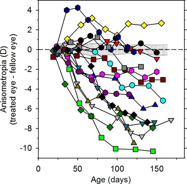
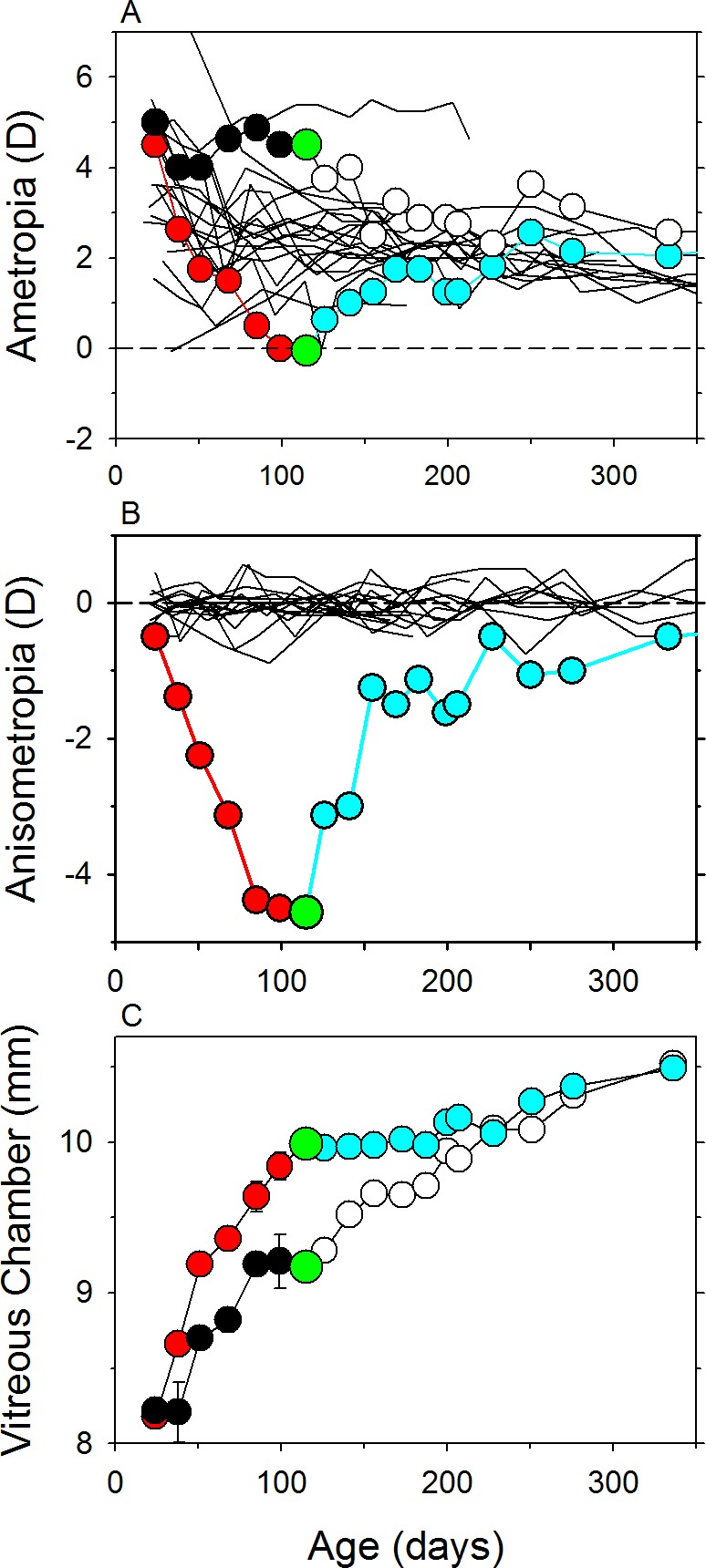
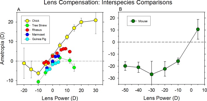
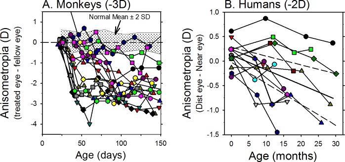
The results of many studies in a variety of species have significantly advanced our understanding of the role of visual experience and the mechanisms of postnatal eye growth, and the development of myopia. This paper surveys and reviews the major contributions that experimental studies using animal models have made to our thinking about emmetropization and development of myopia. These studies established important concepts informing our knowledge of the visual regulation of eye growth and refractive development and have transformed treatment strategies for myopia. Several major findings have come from studies of experimental animal models. These include the eye's ability to detect the sign of retinal defocus and undergo compensatory growth, the local retinal control of eye growth, regulatory changes in choroidal thickness, and the identification of components in the biochemistry of eye growth leading to the characterization of signal cascades regulating eye growth and refractive state. Several of these findings provided the proofs of concepts that form the scientific basis of new and effective clinical treatments for controlling myopia progression in humans. Experimental animal models continue to provide new insights into the cellular and molecular mechanisms of eye growth control, including the identification of potential new targets for drug development and future treatments needed to stem the increasing prevalence of myopia and the vision-threatening conditions associated with this disease.


Figures

Figure 1
Figure 1
Eye growth in experimental animal models. (A) Axial length plotted as a function of age for individual rhesus monkeys., The symbols represent cross-sectional data; the thin black lines represent longitudinal data for individual monkeys. The solid red line shows the best-fitting double exponential function. The horizontal and vertical dashed lines show half-maximum axial length and the age when it was obtained, respectively. (B) Relative axial length changes for different species. The same double exponential function was used to fit the data for each species (humans, black line; rhesus monkey,, red line; tree shrew,, green line; mouse,,,– blue line; guinea pigs,,, cyan line; chicks ,,, pink line; marmoset,, dark red line) and the functions were normalized to the total change in axial length that occurred from birth or eye opening and adulthood. For mice and tree shrews the abscissa represents days of visual experience.
Figure 2
Figure 2
Emmetropization in experimental animal models. The left column shows refractive errors for (A) rhesus monkeys,, (B) marmosets,, (C) tree shews,,, (D) guinea pigs,,, and (E) chicks,,,, plotted as a function of age (or days of visual experience for tree shrews). Longitudinal data from individual animals are shown as solid lines without symbols. Cross-sectional data for individual animals are represented by individual data points. Symbols connected by lines show mean data (typically cross-sectional) from a given study. The solid red lines represent the small eye artifact associated with common measurement techniques like retinoscopy. The middle and right columns contain refractive error frequency distributions obtained near birth/hatching and at ages when refractive development was relatively stable, respectively. The red lines in the histograms show the Gaussian distributions calculated using the mean and standard deviations of the data.
Figure 3
Figure 3
The mouse model of FDM. (A) Mean (±SD) refractive errors plotted as a function of age for C57BL mice.,,,,,– (B) standard deviations of the mean refractive errors from the left panel are plotted as a function of age. The dashed red line represents the best-fitting linear regression and its 95% CIs.
Figure 4
Figure 4
Examples of FDM in animal models and humans. Refractive error frequency distributions for normal (open symbols) and form-deprived eyes (filled symbols) from chicks (A), rhesus monkeys,, (B), and humans (C). Form deprivation was produced in chicks using diffuser lenses; the data were obtained after either 28 or 42 days of age. Form deprivation was produced by surgical eyelid closure in monkeys; the data were obtained over a range of ages and durations of deprivations. Form deprivation in children occurred as a result of conditions (hemangioma and eyelid ptosis) that interfered with a clear retinal image.
Figure 5
Figure 5
The effects of form deprivation are variable. Interocular differences in refractive error (treated eye − fellow eye) plotted as a function of age for individual rhesus macaque monkeys reared with monocular diffuser lenses. The first symbol of each plot represents the onset of form deprivation. The shaded area in each plot represents ±2 SDs of the mean anisometropia for normal control monkeys (adapted from Hung et al.470).
Figure 6
Figure 6
Example of recovery from FDM in rhesus macaques. (A) Spherical-equivalent refractive error plotted as a function of age for the treated (red and cyan symbols) and fellow control eyes (black and white symbols). (B) Interocular differences in refractive error for the same animal plotted as a function of age. (C) Vitreous chamber depth plotted as a function of age for the treated (red and cyan symbols) and fellow control eyes (black and white symbols). The first symbols represent the onset of treatment. The red and black symbols indicate the treatment period. The large green symbols represent the onset of the recovery period. The open and cyan symbols indicate the recovery period. The solid black lines in the top and middle panels are data from untreated control monkeys.
Figure 7
Figure 7
Compensation for lens-imposed retinal defocus occurs in a variety of species (A) chicks, tree shrews, marmosets, rhesus macaques, and guinea pigs, and (B) mice.,, The mean ametropia obtained at the end of the lens-rearing period is plotted as a function of the power of the treatment lenses.
Figure 8
Figure 8
Examples of anisometropic compensation in individual infant rhesus macaque monkeys ([A] adapted from Hung L-F, Arumugam B, She Z, Ostrin L, Smith EL III. Narrow-band, long-wavelength lighting promotes hyperopia and retards vision-induced myopia in infant rhesus monkeys. Exp Eye Res. 2018;176:147–160. Copyright © 2018 Elsevier Ltd.)470 and adolescent humans (age of onset 11 years) ([B] adapted from Phillips JR. Monovision slows juvenile myopia progression unilaterally. Br J Ophthalmol. 2005;89:1196–200. Copyright © 2005 British Journal of Ophthalmology).206 The first symbol in each plot represents the onset of treatment. The monkeys were reared with a −3 D lens in front of their treated eyes and a plano lens in front of their fellow eyes. The human subjects were corrected using a monovision contact lens strategy. The dominant eyes were corrected for distance; the fellow eyes were uncorrected by <2 D.
Figure 9
Figure 9
(A) Chick scleral cross section. The cartilaginous part (cart. sclera) facing the choroid and the fibrous part (fibr. sclera) forming the outer shell can be easily distinguished in this Toluidine blue stained semithin section. (B, C) Electron micrographs of marmoset sclera. (B) Layers of collagen fibers with various orientation are detectable. White arrowhead indicates the cell body of a fibroblast embedded between ECM layers. (C) Higher magnification showing longitudinally (white arrow) and cross-sectional (black arrow) collagen fiber bundles.
Figure 10
Figure 10
Changes in corneal power and anterior chamber depth found in different animal models with experimentally induced myopia. The x- and y-axis parameters represent either interocular difference (treated eye − fellow control eye) or intergroup differences (treated group − normal group). The filled and open symbols represent statistically significant and insignificant changes, respectively. Gray symbols indicate studies that did not perform statistical tests. Numbers inside or near each symbol represent different studies. █ Chicks: (1) Wallman et al., diffusers; (2) Gottlieb et al., diffusers; (3) Hayes et al., diffusers; (4) Irving et al., lenses; (5) Troilo et al., diffusers; (6) Napper et al., diffusers; (7) Napper et al., diffusers; ⧫ tree shrews: (8) Guggenheim et al., diffusers; (9) Siegwart et al., diffusers; (10) McBrien et al., lid-suture; ▴ guinea pigs: (11) Howlett et al., diffusers; ▾ marmosets: (12) Graham and Judge, negative lenses; (13) Troilo and Nickla, diffusers; • rhesus monkeys: (14) Smith and Hung, diffusers; (15) Qiao-Grider et al., diffusers; and (16) Qiao-Grider et al., diffusers and negative lenses, induced myopia was not available, myopic anisometropia of more than −1.0 D was used. For chicks, the corneal radius of curvature values was converted to corneal powers using a refractive index of n' = 1.369.
Figure 11
Figure 11
MRIs obtained in the horizontal plane for the treated (left) and control eyes (right) of rhesus macaque monkeys reared with monocular full-field form deprivation (A) and monocular nasal-field form deprivation (B) (adapted from Smith EL III. Prentice Award Lecture 2010: a case for peripheral optical treatment strategies for myopia. Optom Vis Sci. 2011;88:1029–44. Copyright © 2011 American Academy of Optometry). The nasal and temporal retinas are designated as N and T, respectively. In the middle panels, the outlines for the treated (red) and fellow eyes (blue) have been superimposed after rotating the fellow eye images around the optic axes so that the nasal retinas (N) are shown to the right for both eyes. The superimposed images were aligned using the lines that connected the equatorial poles of the crystalline lenses as a reference (the red lines shown in the treated- and fellow-eye images in the left and middle columns).
Figure 12
Figure 12
Longitudinal changes in spherical equivalent refractive errors for the right eyes of infant rhesus macaque monkeys reared with binocular −3 D lenses. The monkeys represented in panel A wore the lenses continuously throughout the daily 12-hour lights-on cycle. For the monkeys represented in panel B, the −3 D lenses were removed for four 15-minute periods during the daily 12-hour lights-on cycle. The black lines in the upper plots show data from normal infant monkeys. The schematic in the lower left (C) shows the times when these animals were allowed unrestricted vision. The lower right plot (D) compares plotted as mean end-of-treatment ametropias for normal monkeys and the two experimental groups of monkeys (adapted from Kee C-S, Hung L-F, Qiao-Grider Y, et al. Temporal constraints on experimental emmetropization in infant monkeys. Invest Ophthalmol Vis Sci. 2007;48:957–962. Copyright © 2007 The Association for Research in Vision and Ophthalmology, Inc.).
Figure 13
Figure 13
Effects of multifocal lens rearing. (A) Comparisons of the effects of dual focus, Fresnel-like lenses (50:50 area ratios) on refractive error development in rhesus macaques, chicks, marmosets, and guinea pigs. The left scale indicates the relative percentage change in ametropias at the end of treatment. For binocularly treated animals (rhesus monkeys), the ametropias for the right eyes are represented relative to that for control animals. For monocularly treated animals (all other species), the ametropias for the treated eyes are expressed relative to that of the fellow eye. Values of 0% and 100% indicate complete compensation for the most hyperopic and myopic image planes, respectively. Values of 50% indicate that the animals compensated for the average power of the dual focus treatment lenses (adapted from Arumugam B, Hung L-F, To C-H, Holden B, Smith EL III. The effects of simultaneous dual focus lenses on refractive development in infant monkeys. Invest Ophthalmol Vis Sci. 2014;55:7423–7432. Copyright © 2014 The Association for Research in Vision and Ophthalmology, Inc.). (B) The average ametropias for infant rhesus monkeys reared with dual focus Fresnel lenses (either +3 D and plano or −3 D and plano) plotted as a function of the percentage of surface areas that was devoted to the powered portions of the treatment lenses. Control monkeys reared with unrestricted vision are represented at the 0 point on the abscissa. Control monkeys reared with −3 and +3 D single-vision lenses are represented at the “100% −3 D” and “100% +3 D” positions, respectively. The dual-focus groups are positioned according to the proportion of lens surface areas devoted to the −3 and +3 D power zones (adapted from Arumugam B, Hung L-F, To C-H, Sankaridurg P, Smith EL III. The effects of the relative strength of simultaneous competing defocus signals on emmetropization in infant rhesus monkeys. Invest Ophthalmol Vis Sci. 2016;57:3949–3960. Licensed under a Creative Commons Attribution-NonCommercial-NoDerivatives 4.0 International License.).323
Figure 14
Figure 14
The effects of imposing defocus on the peripheral retina. (A, B) Spherical equivalent refractive corrections obtained at ages corresponding to the end of the lens-rearing period for control monkeys (open diamonds) and monkeys reared with either diffusers ([A] adapted from Smith EL III, Kee C-S, Ramamirtham R, Qiao-Grider Y, Hung L-F. Peripheral vision can influence eye growth and refractive development in infant monkeys. Invest Ophthalmol Vis Sci. 2005;46:3965–3972. Copyright © 2005 The Association for Research in Vision and Ophthalmology, Inc.) or −3 D lenses ([B] adapted from Smith EL III, Hung LF, Huang J. Relative peripheral hyperopic defocus alters central refractive development in infant monkeys. Vis Res. 2009;49:2386–2392. Copyright © 2009 Elsevier Ltd.). The solid green and red symbols represent monkeys that worn treatment lenses that had central apertures that provided unrestricted vision for the central 24° to 32°. For comparison purposes, the half-filled diamonds represent monkeys that were reared with intact diffusers or −3 D lenses that altered vision across the entire field. The horizontal dashed line represents the average refractive error for the control monkeys; the solid lines denote ±1 SD from the control mean. (C) Changes in refractive error produced by rearing chicks with +5 D treatment lenses that had varying diameter central apertures that allowed unrestricted central vision (adapted from Liu Y, Wildsoet C. The effect of two-zone concentric bifocal spectacle lenses on refractive error development and eye growth in young chicks. Invest Ophthalmol Vis Sci. 2011;52:1078–1086. Copyright © 2011 Association for Research in Vision and Ophthalmology).
Figure 15
Figure 15
Emmetropization and experimentally altered refractive state in tree shrew. (Top) Refraction as a function of days after eye opening (days of visual experience, or DVE). Each line is for an individual animal. Data for untreated animals and red light are shown as the average of both eyes; data for −5 D lens animals are for the treated eye only. Untreated tree shrews were raised under fluorescent colony lighting (data from Gawne et al.467), red light animals were exposed to ambient narrow-band red light stating at 95 DVE, and −5 D lens animals wore a monocular −5 D lens over one eye. Binocular +5 D lenses were worn starting at either 11 or 24 DVE. (Bottom) Axial length of the eyes as a function of time for the normal animals shown in the top panel.
Figure 16
Figure 16
Does a biochemical signal cascade beginning in retina and ending with changes in sclera extracellular matrix control eye growth and refractive state? Biochemical retinal signal(s) in response to myopic or hyperopic defocus conditions, may initiate a signal pathway cascade from retina to RPE, the choroid, and eventually sclera, controlling the remodeling and synthesis of scleral extracellular matrix and eye growth directly or in response to IOP effects. A general list of substances and functions implicated at different steps in experimentally induced changes in eye growth and refractive state are indicated. PG, proteoglycan; GAG, glycosaminoglycans.
Figure 17
Figure 17
Information flow from myopiagenic stimuli signals that would produce gene expression changes related to signaling, degradative enzymes, and inhibitors and ECM proteins (adapted from Guo L, Frost MR, He L, Siegwart JT Jr, Norton TT. Gene expression signatures in tree shrew sclera in response to three myopiagenic conditions. Invest Ophthalmol Vis Sci. 2013;54:6806–6819. Copyright © 2013 Association for Research in Vision and Ophthalmology).
Figure 18
Figure 18
A heuristic model of the visually regulated control of eye growth and refractive state.

References

    1. Holden BA, Fricke TR, Wilson DA, et al. Global prevalence of myopia and high myopia and temporal trends from 2000 through 2050. Ophthalmology. 2016;123:1036–1042. - PubMed
    1. Turnbull PR, Backhouse S, Phillips JR. Visually guided eye growth in the squid. Curr Biol. 2015;25:R791–R792. - PubMed
    1. Jeon CJ, Strettoi E, Masland RH. The major cell populations of the mouse retina. J Neurosci. 1998;18:8936–8946. - PMC - PubMed
    1. Wu SM. From retinal circuitry to eye diseases – in memory of Henk Spekreijse. Vision Res. 2009;49:992–995. - PMC - PubMed
    1. Walls GL. The Vertebrate Eye and its Adaptive Radiations Hafner Publishing 1970 ed. Bloomfield Hills: Cranbrook Institute of Science;; 1942.

Publication types