. 2019 Jul;40(7):908-925.
doi: 10.1002/humu.23731. Epub 2019 Apr 24.

SLC35A2-CDG: Functional characterization, expanded molecular, clinical, and biochemical phenotypes of 30 unreported Individuals

Bobby G Ng  1 Paulina Sosicka  1 Satish Agadi  2 Mohammed Almannai  2 Carlos A Bacino  2   3 Rita Barone  4   5 Lorenzo D Botto  6 Jennifer E Burton  7 Colleen Carlston  8 Brian Hon-Yin Chung  9 Julie S Cohen  10 David Coman  11   12 Katrina M Dipple  13   14   15 Naghmeh Dorrani  16 William B Dobyns  17   18 Abdallah F Elias  19 Leon Epstein  20 William A Gahl  21   22 Domenico Garozzo  5 Trine Bjørg Hammer  23 Jaclyn Haven  19 Delphine Héron  24 Matthew Herzog  15 George E Hoganson  7 Jesse M Hunter  25 Mahim Jain  10 Jane Juusola  26 Shenela Lakhani  27 Hane Lee  15   28 Joy Lee  29   30 Katherine Lewis  11 Nicola Longo  6 Charles Marques Lourenço  31 Christopher C Y Mak  9 Dianalee McKnight  26 Bryce A Mendelsohn  32 Cyril Mignot  24 Ghayda Mirzaa  17   18 Wendy Mitchell  33   34 Hiltrud Muhle  35 Stanley F Nelson  15   28   36 Mariusz Olczak  37 Christina G S Palmer  15   36   38 Arthur Partikian  39 Marc C Patterson  40 Tyler M Pierson  41   42   43 Shane C Quinonez  44 Brigid M Regan  45 M Elizabeth Ross  27 Maria J Guillen Sacoto  26 Fernando Scaglia  2   3   46 Ingrid E Scheffer  45   47 Devorah Segal  27   48 Nilika Shah Singhal  49 Pasquale Striano  50 Luisa Sturiale  5 Joseph D Symonds  51 Sha Tang  25 Eric Vilain  52 Mary Willis  53 Lynne A Wolfe  21   22 Hui Yang  26 Shoji Yano  54 Zöe Powis  25 Sharon F Suchy  25 Jill A Rosenfeld  26 Andrew C Edmondson  2 Stephanie Grunewald  55 Hudson H Freeze  56
Affiliations

SLC35A2-CDG: Functional characterization, expanded molecular, clinical, and biochemical phenotypes of 30 unreported Individuals

Bobby G Ng et al. Hum Mutat. 2019 Jul.

Abstract

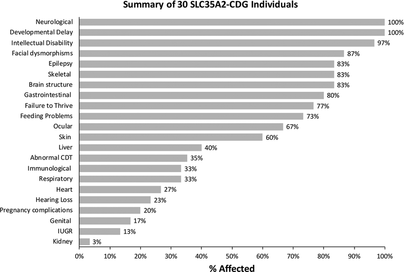
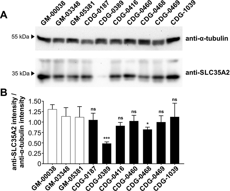
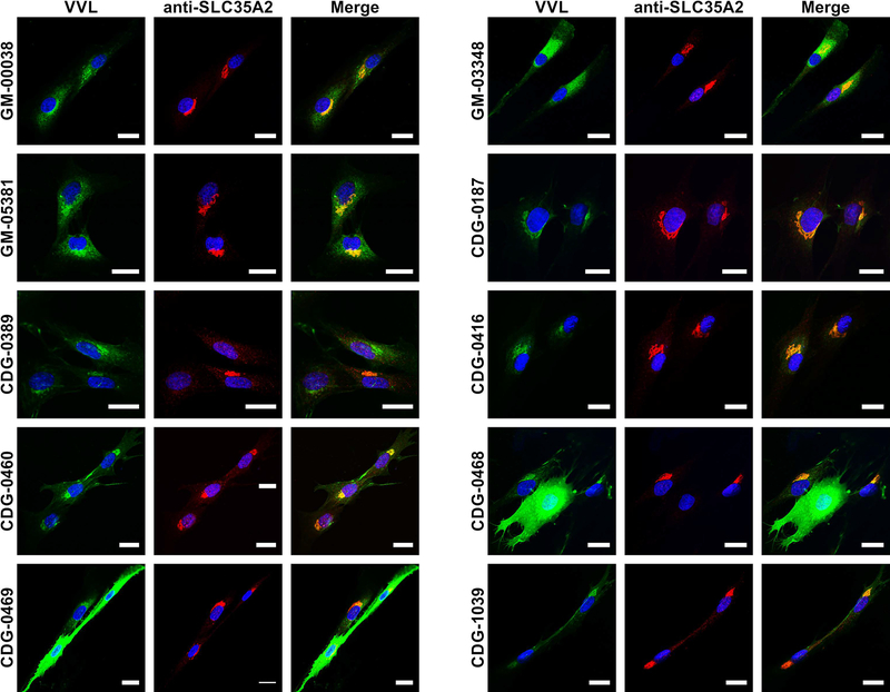
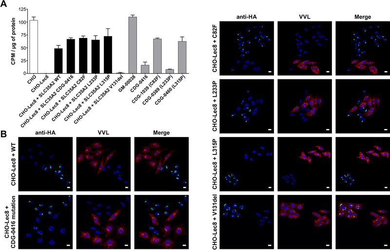
Pathogenic de novo variants in the X-linked gene SLC35A2 encoding the major Golgi-localized UDP-galactose transporter required for proper protein and lipid glycosylation cause a rare type of congenital disorder of glycosylation known as SLC35A2-congenital disorders of glycosylation (CDG; formerly CDG-IIm). To date, 29 unique de novo variants from 32 unrelated individuals have been described in the literature. The majority of affected individuals are primarily characterized by varying degrees of neurological impairments with or without skeletal abnormalities. Surprisingly, most affected individuals do not show abnormalities in serum transferrin N-glycosylation, a common biomarker for most types of CDG. Here we present data characterizing 30 individuals and add 26 new variants, the single largest study involving SLC35A2-CDG. The great majority of these individuals had normal transferrin glycosylation. In addition, expanding the molecular and clinical spectrum of this rare disorder, we developed a robust and reliable biochemical assay to assess SLC35A2-dependent UDP-galactose transport activity in primary fibroblasts. Finally, we show that transport activity is directly correlated to the ratio of wild-type to mutant alleles in fibroblasts from affected individuals.

Keywords: UDP-galactose; congenital disorders of glycosylation; glycoside; nucleotide sugar transporter.


Conflict of interest statement

CONFLICT OF INTEREST STATEMENT

The Department of Molecular and Human Genetics at Baylor College of Medicine derives revenue from clinical genetic testing completed at Baylor Genetics Laboratories. Fernando Scaglia is involved in clinical trials supported by BioElectron, Reata Pharmaceuticals, and Stealth BioTherapeutics. Mary Willis declares views expressed herein are those of the authors and do not necessarily reflect the official policy or position of the Department of the Navy, Department of Defense, or the U.S. Government. Zöe Powis, Jesse M. Hunter, Sha Tang are employed by Ambry Genetics. Sharon F. Suchy, Jane Juusola, Dianalee McKnight, Maria Guillen Sacato and Hui Yang are employed by GeneDx, Inc., a wholly-owned subsidiary of OPKO Health, Inc. The other authors have no conflict.

Figures

Figure 1.
Figure 1.
Schematic representation of the SLC35A2 variants present in our 30 individuals. (A) Schematic representation showing exon location of all 30 SLC35A2 variants identified in this study. (B) Schematic representation showing protein localization of all 30 SLC35A2 variants identified in this study. SLC35A2 topology was determined using 3D model generated with PHYRE2 server (Kelley, Mezulis, Yates, Wass & Sternberg, 2015).
Figure 2.
Figure 2.
Clinical summary of 30 individuals with de novo variants in SLC35A2. Clinical information for each of the 30 individuals were provided by clinicians and summarized as a percentage of affected individuals.
Figure 3.
Figure 3.
Optimization of the glycoside-based UDP-galactose transport assay. (A) GlcNAcβ−4-MU concentration, temperature and time of the assay were optimized. Glycoside concentration was tested using CHO (circles) and CHO-Lec8 (squares) cells. Temperature and time were optimized only on CHO lines. Transport efficiency at room temperature (diamonds) and 37 °C (triangles) was compared using increasing concentrations of GlcNAcβ−4-MU. Time curve (asterisk) was assayed at 37°C with 1 mM glycoside. (B) UDP-galactose transport was measured in CHO cells using optimized conditions. As a negative control CHO-Lec8 cells were employed. Specificity of the assay was determined by treating CHO cells with 0.025% Trition-X100 (CHO + TX100). The assay was performed in three biological repetitions. Error bars represent SD (C) Michaelis constant and the maximum UDP-galactose transport velocity were determined for CHO protein in optimized assay conditions. Each point represents a mean of 3 to 6 independent biological repetitions using different cell preparations and performed on different days. Error bars represents SEM. CPM – Counts Per Minute
Figure 4.
Figure 4.
UDP-galactose transport assay in primary fibroblasts. UDP-[6-3H] galactose transport in permeabilized fibroblast cells with intact Golgi apparatus under optimized conditions. Each assay was performed in three biological repetitions and expressed as a percent of the control (* p < 0.05; ** p < 0.005; *** p<0.001).
Figure 5.
Figure 5.
Determination of the wild-type to mutant allele ratios in fibroblasts (A) SLC35A2 cDNA allele ratios in primary fibroblasts from control and SLC35A2-CDG subjects. Wild-type to mutant allele ratios were determined using a restriction enzyme-based assay. GM-00038, GM-03348 and GM-05381 cells were used as controls. Each assay was performed in duplicate in order to determine reproducibility of the assay. (B) UDP-galactose transporter activity was calculated based on allele ratio presented as a percentage of GM-00038 transport activity. To illustrate the activity of the respective mutant protein, the contribution of wild-type protein was subtracted.
Figure 6.
Figure 6.
Western Blot analysis of SLC35A2 protein from primary fibroblast. (A) Western Blot analysis of SLC35A2 protein from a total of seven SLC35A2-CDG fibroblast lines as well as three controls (GM-00038, GM-03348 and GM-05381). Anti-α-tubulin antibody was used as a loading control. (B) Quantification was performed by normalizing SLC35A2 to α-tubulin from four independent blots using two biological replicates collected on separate days with error bars calculated as a StdDev.
Figure 7.
Figure 7.
Immunofluorescence staining of primary fibroblasts. To assess glycosylation between control (GM-00038, GM-03348 and GM-05381) and subject fibroblasts (CDG-0187, CDG-0389, CDG-0416, CDG-0460, CDG-0468, CDG-0469 and CDG-1039) VVL lectin was used (green). This lectin specifically recognizes terminal GalNAc present in O-glycans, which is usually masked by galactose and blocks the lectin reactivity. UDP-galactose transporter was counterstained with anti-SLC35A2 antibody (red). Scale bar 20 μm.
Figure 8.
Figure 8.
Characterization of CHO-Lec8 stable clones expressing SLC35A2. (A) UDP-[6-3H] galactose transport assay in parental CHO, CHO-Lec8 and CHO-Lec8 cells overexpressing respective variants of SLC35A2 protein. For better comparison, fibroblast results from control (GM-00038) and four affected individuals (CDG-0389, CDG-0416, CDG-0460, CDG-1039) were plotted. The transport for SLC35A2-CDG fibroblasts was expressed as the predicted activity of mutant transporter by subtracting the contribution of wild type protein, based on the calculated allele ratio. Each assay was performed in two biological repetitions. (B) Immunofluorescence staining of CHO-Lec8 cells overexpressing UDP-galactose transporter. Overexpressed HA-tagged SLC35A2 protein was detected with anti-HA antibody (green). VVL lectin staining was used to detect glycosylation changes between CHO-Lec8 cells which do or do not overexpress the transporter (red). Stable transfectants, but not clonal populations were analyzed. Scale bar 20 μm.

References

    1. Allen RC, Zoghbi HY, Moseley AB, Rosenblatt HM, & Belmont JW (1992). Methylation of HpaII and HhaI sites near the polymorphic CAG repeat in the human androgen-receptor gene correlates with X chromosome inactivation. Am J Hum Genet, 51(6), 1229–1239. - PMC - PubMed
    1. Bosch DG, Boonstra FN, de Leeuw N, Pfundt R, Nillesen WM, de Ligt J, … de Vries BB (2016). Novel genetic causes for cerebral visual impairment. Eur J Hum Genet, 24(5), 660–665. doi: 10.1038/ejhg.2015.186 - DOI - PMC - PubMed
    1. Brandli AW, Hansson GC, Rodriguez-Boulan E, & Simons K (1988). A polarized epithelial cell mutant deficient in translocation of UDP-galactose into the Golgi complex. J Biol Chem, 263(31), 16283–16290. - PubMed
    1. Brockhausen I, & Stanley P (2015). O-GalNAc Glycans In rd, Varki A, Cummings RD, Esko JD, Stanley P, Hart GW, Aebi M, Darvill AG, Kinoshita T, Packer NH, Prestegard JH, Schnaar RL, & Seeberger PH (Eds.), Essentials of Glycobiology (pp. 113–123). Cold Spring; Harbor (NY).
    1. Bruneel A, Cholet S, Drouin-Garraud V, Jacquemont ML, Cano A, Megarbane A, … Fenaille F (2018). Complementarity of electrophoretic, mass spectrometric, and gene sequencing techniques for the diagnosis and characterization of congenital disorders of glycosylation. Electrophoresis. doi: 10.1002/elps.201800021 - DOI - PubMed

Publication types

Substances