. 2019 Jul;24(6):615-627.
doi: 10.1177/2472555219830087. Epub 2019 Feb 28.

3D Cell-Based Assays for Drug Screens: Challenges in Imaging, Image Analysis, and High-Content Analysis

Affiliations

3D Cell-Based Assays for Drug Screens: Challenges in Imaging, Image Analysis, and High-Content Analysis

Tijmen H Booij et al. SLAS Discov. 2019 Jul.

Abstract

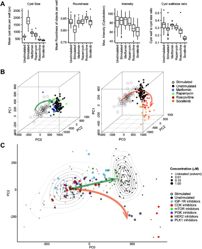
The introduction of more relevant cell models in early preclinical drug discovery, combined with high-content imaging and automated analysis, is expected to increase the quality of compounds progressing to preclinical stages in the drug development pipeline. In this review we discuss the current switch to more relevant 3D cell culture models and associated challenges for high-throughput screening and high-content analysis. We propose that overcoming these challenges will enable front-loading the drug discovery pipeline with better biology, extracting the most from that biology, and, in general, improving translation between in vitro and in vivo models. This is expected to reduce the proportion of compounds that fail in vivo testing due to a lack of efficacy or to toxicity.

創薬の前臨床早期において、ハイコンテントイメージング自動アナリシスを併用しての適性度の高い細胞モデルの導入は、新薬開発パイプラインの前臨床段階に進む化合物の質を高めるものと期待される。本レビューでは、ハイスループットスクリーニングおよびハイコンテントアナリシスにおける、適性度の高い三次元細胞培養モデルへの現行の切り替えとそれに伴う課題について検討する。それらの課題を克服することで、適切なバイオロジーによる創薬パイプラインのフロントローディングが可能になり、そのバイオロジーから最大限のものを引き出し、概してin vivo外挿(translation between in vitro and in vivo models)を向上させることを我々は提案する。これにより、有効性欠如または毒性が原因でin vivo試験に不合格となる化合物の割合を低下できるものと期待される。

대용량 이미징과 자동화 분석과 함께 초기 임상 전 신약 개발에서 관련성이 더 높은 세포 모형을 도입하면 신약 개발에서 임상 전 단계로 넘어가는 물질의 품질을 높여줄 것으로 기대된다. 본 고찰에서는 관련성이 더 높은 3D 세포 배양 모형으로 넘어가고 있는 현재의 변화와 이에 따른 초고속 스크리닝 및 대용량 분석의 문제점을 논하고자 한다. 이러한 문제점을 극복하면 신약 개발의 초반 부분에 더 나은 세포 작용을 제공하고, 이러한 세포 작용에서 최대한 추출을 하고, 세포 및 동물 모형 사이의 중개를 개선시킬 것이라 제안한다. 이는 효능과 독성 때문에 동물 시험에서 실패하는 물질의 비율을 감소시킬 것으로 기대된다.

在早期临床前药物研发中引入相关性更好的细胞模型,结合高内涵成像和自动化分析,有望提高药物开发过程中进展至临床前阶段的化合物的质量。在本综述中,我们讨论了目前相关性更好的三维细胞培养模型的应用情况,以及相应高通量筛选和高内涵分析种的挑战。我们认为,克服这些挑战可以为药物研发流程提供更好的前期生物学基础,最大化的利用这些生物学基础,并在总体上改善从体外模型到体内模型的转换。预计这将减少因药效缺乏或毒性而在体内测试中失败的化合物的比例。

Keywords: 3D culture; 3D 배양; high-content screening; image analysis; imaging; イメージアナリシス; イメージング; ハイコンテントスクリーニング; 三次元細胞培養; 三维细胞培养; 关键词 高内涵筛选; 图像分析; 成像; 대용량 스크리닝; 이미지 분석; 이미징.


Conflict of interest statement

Declaration of Conflicting Interests: The authors disclosed the following potential conflicts of interest with respect to the research, authorship, and/or publication of this article: Leo S. Price is founder and shareholder of the compound testing CRO OcellO B.V.

Figures

Figure 1.
Figure 1.
3D cell cultures provide a more physiologically relevant context for drug screening. (A) Prostate carcinoma (PC-3) cells cultured as 2D monolayer (top) show negligible morphological changes in response to growth factor (20 ng/mL hrEGF) stimulation but become invasive if embedded in 3D hydrogels (bottom) after growth factor stimulation. These invasive characteristics can be used to investigate the efficacy of inhibitors of receptor tyrosine kinases. Images in the top panel were obtained using a wide-field BD pathway 855 with a 10× objective, and images in the bottom panel were obtained using a Nikon Ti Eclipse confocal microscope with a 20× objective. (B) mIMCD3 cells transduced with a short-hairpin targeting Pkd1 form a monolayer in 2D culture (left panel, BD pathway 855 with 10× objective), but form cysts in 3D hydrogels, representing a more pathophysiologically relevant model of PKD (right panel, Nikon Ti Eclipse confocal microscope with 20× objective). F-actin (rhodamine-phalloidin), red; nuclei (Hoechst 33258), blue.
Figure 2.
Figure 2.
Nomenclature of 3D cell-based assays.
Figure 3.
Figure 3.
3D cultures of PDX material. PDX material from different tumors can be cultured in 3D hydrogels to form complex microtissues that can be used for compound screening in a preclinically relevant context. Actin cytoskeleton visualized with rhodamine-phalloidin. PDX tumor material provided by Charles River Labs (Freiburg, Germany). Annotations refer to tumor type and PDX model number; BX = bladder; MAX = mammary; GX = gastric; PAX = pancreatic; LX = lung. 3D cultures and images generated by OcellO B.V.
Figure 4.
Figure 4.
Maximum-intensity projections can cause loss of phenotypic information in 3D cultures. (A) Schematic representation of 2D maximum-intensity projections modified from Booij et al. (2016). Structures embedded in hydrogels are captured in xy and z directions using automated microscopy, and in-focus regions from all sections are projected into a 2D reconstruction. (B) 2D projections from 3D structures can cause loss of important phenotypic characteristics. These images display human kidney cyst-derived organoids, cultured in Matrigel and stained for F-actin (rhodamine-phalloidin, red) and nuclei (Hoechst 33258, blue) and imaged on a Nikon Ti Eclipse confocal microscope. Example 3D reconstructions from image sections obtained after imaging with a Nikon Ti Eclipse confocal microscope are included as Supplemental Video 1. Maximum-intensity projection performed with ImageJ software prevents lumen and cell shape detection.
Figure 5.
Figure 5.
Exploiting multiparametric data to discriminate responses. (A) Metformin, rapamycin, roscovitine, and sorafenib inhibit forskolin-stimulated PKD cyst swelling, with roscovitine and sorafenib inducing the most potent response based on evaluation of individual parameters. Analysis was performed using Ominer software (OcellO B.V.). All displayed phenotypic parameters are derived from the rhodamine-phalloidin (f-actin) staining of 3D-cultured cysts. (B) Left panel: Three principal components summarizing 84% of variance in the data show a desirable phenotypic change (green arrow) in which 5 mM metformin (blue) and 10 nM rapamycin (green) revert a 2.5 µM forskolin-stimulated phenotype (swollen cyst, empty circles) to one indistinguishable from an unstimulated (solvent) phenotype (solid black circles). Right panel: 31.6 µM roscovitine and 10 µM sorafenib induce an aberrant phenotype (orange arrow); data points represent single wells. Figures adapted from Booij et al. (2017). (C) Two principal components from B showing multiple inhibitors targeting cyclin-dependent kinases (CDK), mammalian target of rapamycin (mTOR), phosphatidylinositol-4,5-bisphosphate 3-kinase (PI3K), human epidermal growth factor receptor 2 (HER2), and polo-like kinase (PLK1). Contour plots represent density estimations to emphasize the locations of forskolin-stimulated (empty circles) and unstimulated controls (solid black circles), respectively. Green arrow represents desirable compound efficacy, from forskolin-stimulated control (empty circles, swollen cysts) to unstimulated control (solid black circles, small cysts), and the orange arrow represents aberrant phenotypes that are observed after treatment with PLK1 inhibitors or high-dose CDK inhibitors, indicative of cytotoxicity.

References

    1. Langhans S. A. Three-Dimensional In Vitro Cell Culture Models in Drug Discovery and Drug Repositioning. Front. Pharmacol. 2018, 9, 6. - PMC - PubMed
    1. Zheng W., Thorne N., McKew J. C. Phenotypic Screens as a Renewed Approach for Drug Discovery. Drug Discov. Today 2013, 18, 1067–1073. - PMC - PubMed
    1. Swinney D. C., Anthony J. How Were New Medicines Discovered? Nat. Rev. Drug Discov. 2011, 10, 507–519. - PubMed
    1. Swinney D. C., Phenotypic vs. Target-Based Drug Discovery for First-in-Class Medicines. Clin. Pharmacol. Ther. 2013, 93, 299–301. - PubMed
    1. Singh S., Carpenter A. E., Genovesio A. Increasing the Content of High-Content Screening: An Overview. J. Biomol. Screen. 2014, 19, 640–650. - PMC - PubMed

Publication types

MeSH terms

Substances

LinkOut - more resources