

Identification of a mechanogenetic link between substrate stiffness and chemotherapeutic response in breast cancer
- PMID: 30818087
- PMCID: PMC6474249
- DOI: 10.1016/j.biomaterials.2019.02.018
Identification of a mechanogenetic link between substrate stiffness and chemotherapeutic response in breast cancer
Abstract
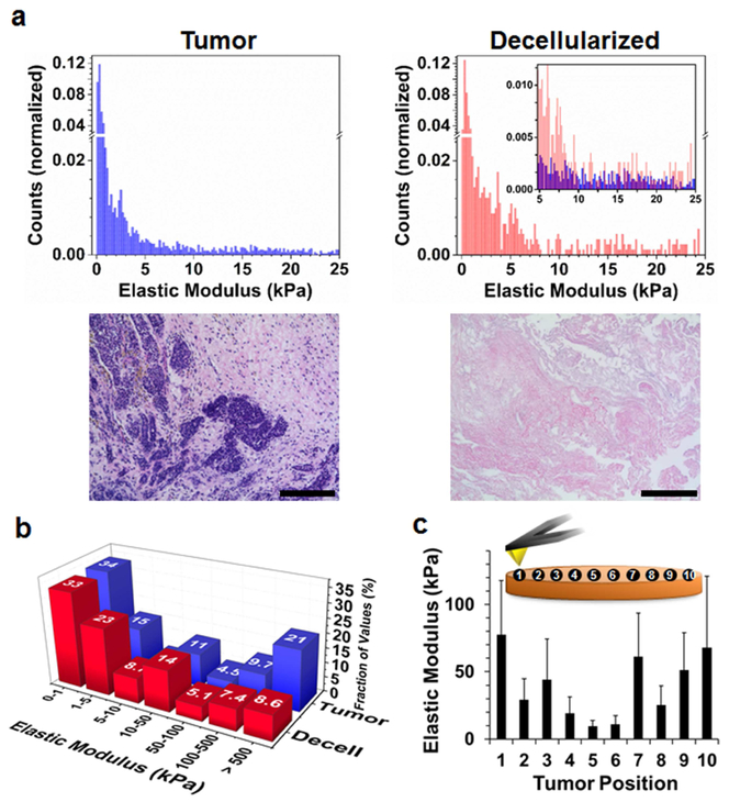
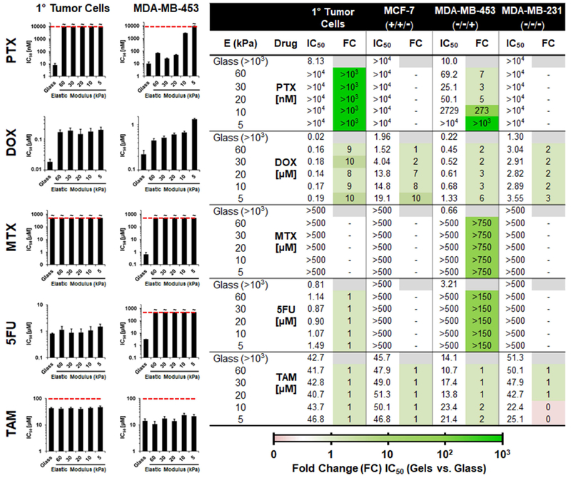
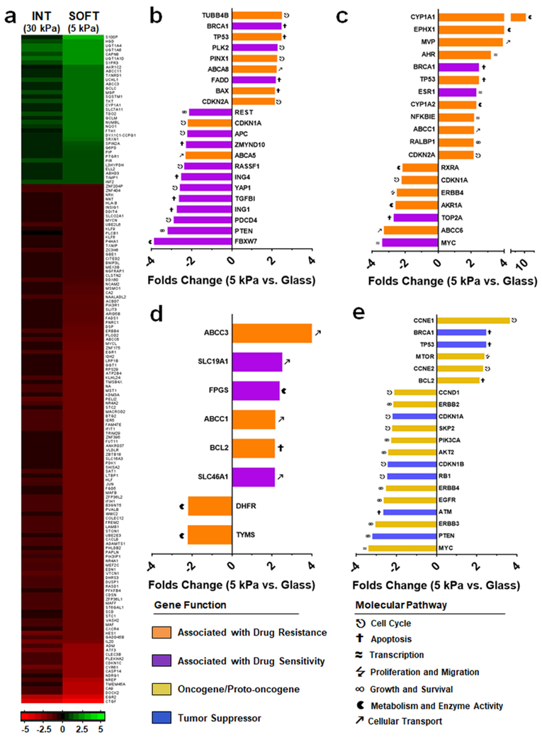
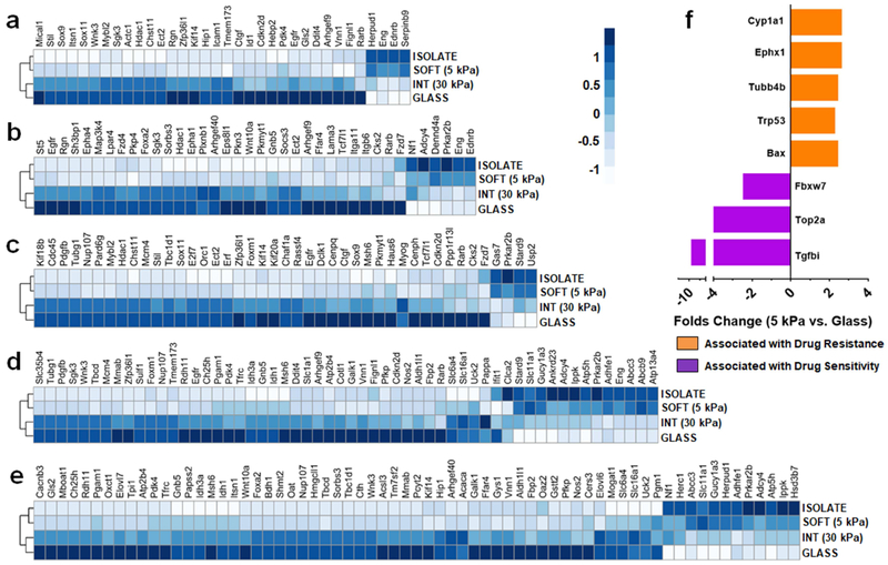
Mechanical feedback from the tumor microenvironment regulates an array of processes underlying cancer biology. For example, increased stiffness of mammary extracellular matrix (ECM) drives malignancy and alters the phenotypes of breast cancer cells. Despite this link, the role of substrate stiffness in chemotherapeutic response in breast cancer remains unclear. This is complicated by routine culture and adaptation of cancer cell lines to unnaturally rigid plastic or glass substrates, leading to profound changes in their growth, metastatic potential and, as we show here, chemotherapeutic response. We demonstrate that primary breast cancer cells undergo dramatic phenotypic changes when removed from the host microenvironment and cultured on rigid surfaces, and that drug responses are profoundly altered by the mechanical feedback cells receive from the culture substrate. Conversely, primary breast cancer cells cultured on substrates mimicking the mechanics of their host tumor ECM have a similar genetic profile to the in situ cells with respect to drug activity and resistance pathways. These results suggest substrate stiffness plays a significant role in susceptibility of breast cancer to clinically-approved chemotherapeutics, and presents an opportunity to improve drug discovery efforts by integrating mechanical rigidity as a parameter in screening campaigns.
Keywords: Chemotherapy; Drug screening; Genotyping; Hydrogel; Mechanobiology.
Copyright © 2019. Published by Elsevier Ltd.
Figures

Similar articles
-
Biofabrication of Tunable 3D Hydrogel for Investigating the Matrix Stiffness Impact on Breast Cancer Chemotherapy Resistance.ACS Biomater Sci Eng. 2025 Mar 10;11(3):1417-1431. doi: 10.1021/acsbiomaterials.4c01636. Epub 2025 Feb 27. ACS Biomater Sci Eng. 2025. PMID: 40013911
-
Distinct phenotypes of cancer cells on tissue matrix gel.Breast Cancer Res. 2020 Jul 31;22(1):82. doi: 10.1186/s13058-020-01321-7. Breast Cancer Res. 2020. PMID: 32736579 Free PMC article.
-
Matrix Stiffness-Regulated Growth of Breast Tumor Spheroids and Their Response to Chemotherapy.Biomacromolecules. 2021 Feb 8;22(2):419-429. doi: 10.1021/acs.biomac.0c01287. Epub 2020 Nov 2. Biomacromolecules. 2021. PMID: 33136364
-
Matrix Rigidity Controls Epithelial-Mesenchymal Plasticity and Tumor Metastasis via a Mechanoresponsive EPHA2/LYN Complex.Dev Cell. 2020 Aug 10;54(3):302-316.e7. doi: 10.1016/j.devcel.2020.05.031. Epub 2020 Jun 22. Dev Cell. 2020. PMID: 32574556 Free PMC article.
-
Mechanical factors in the breast cancer microenvironment: Emphasizing functional adaptation.Biochem Biophys Res Commun. 2025 Jul 22;771:152048. doi: 10.1016/j.bbrc.2025.152048. Epub 2025 May 19. Biochem Biophys Res Commun. 2025. PMID: 40412051 Review.
Cited by
-
Mechanobiology of Dental Pulp Cells.Cells. 2024 Feb 21;13(5):375. doi: 10.3390/cells13050375. Cells. 2024. PMID: 38474339 Free PMC article. Review.
-
Mitochondrial fission links ECM mechanotransduction to metabolic redox homeostasis and metastatic chemotherapy resistance.Nat Cell Biol. 2022 Feb;24(2):168-180. doi: 10.1038/s41556-022-00843-w. Epub 2022 Feb 14. Nat Cell Biol. 2022. PMID: 35165418 Free PMC article.
-
Self-Assembling Polypeptide Hydrogels as a Platform to Recapitulate the Tumor Microenvironment.Cancers (Basel). 2021 Jun 30;13(13):3286. doi: 10.3390/cancers13133286. Cancers (Basel). 2021. PMID: 34209094 Free PMC article.
-
The Extracellular Matrix and Vesicles Modulate the Breast Tumor Microenvironment.Bioengineering (Basel). 2020 Oct 11;7(4):124. doi: 10.3390/bioengineering7040124. Bioengineering (Basel). 2020. PMID: 33050609 Free PMC article. Review.
-
Effect of Electric Fields on the Mechanical Mechanism of Regorafenib-VEGFR2 Interaction to Enhance Inhibition of Hepatocellular Carcinoma.Biomolecules. 2025 Jan 1;15(1):42. doi: 10.3390/biom15010042. Biomolecules. 2025. PMID: 39858437 Free PMC article.
References
-
- Paszek MJ, Zahir N, Johnson KR, Lakins JN, Rozenberg GI, Gefen A, Reinhart-King CA, Margulies SS, Dembo M, Boettiger D, Hammer DA, Weaver VM, Tensional homeostasis and the malignant phenotype, Cancer Cell 8(3) (2005) 241–254. - PubMed
-
- Chaudhuri O, Koshy ST, Branco da Cunha C, Shin J-W, Verbeke CS, Allison KH, Mooney DJ, Extracellular matrix stiffness and composition jointly regulate the induction of malignant phenotypes in mammary epithelium, Nat. Mater. 13(10) (2014) 970–978. - PubMed
Publication types
MeSH terms
Substances
Grants and funding
LinkOut - more resources
Full Text Sources
Other Literature Sources
Molecular Biology Databases
