. 2019 Feb 27;10(3):165.
doi: 10.3390/mi10030165.

Engineering Microfluidic Organoid-on-a-Chip Platforms

Affiliations

Engineering Microfluidic Organoid-on-a-Chip Platforms

Fang Yu et al. Micromachines (Basel). .

Abstract

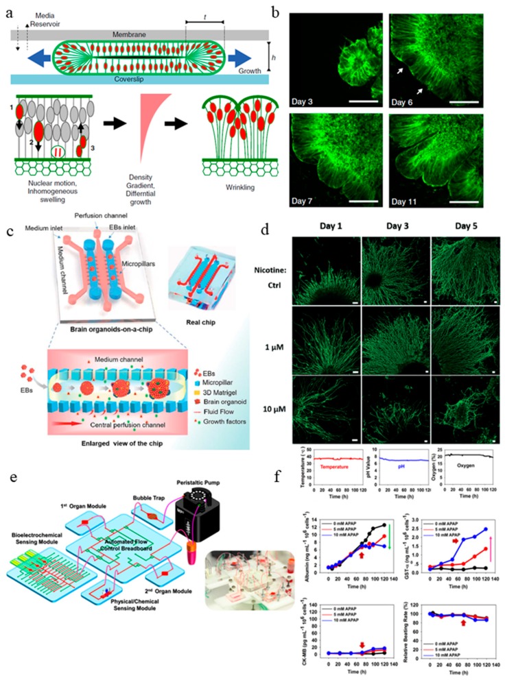
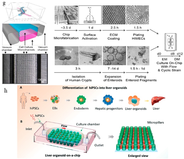
In vitro cell culture models are emerging as promising tools to understand human development, disease progression, and provide reliable, rapid and cost-effective results for drug discovery and screening. In recent years, an increasing number of in vitro models with complex organization and controlled microenvironment have been developed to mimic the in vivo organ structure and function. The invention of organoids, self-organized organ-like cell aggregates that originate from multipotent stem cells, has allowed a whole new level of biomimicry to be achieved. Microfluidic organoid-on-a-chip platforms can facilitate better nutrient and gas exchange and recapitulate 3D tissue architecture and physiology. They have the potential to transform the landscape of drug development and testing. In this review, we discuss the challenges in the current organoid models and describe the recent progress in the field of organoid-on-a-chip.

Keywords: cell culture; drug screening; microbioreactor; microfluidics; organ-on-a-chip; organoids.


Conflict of interest statement

The authors indicated no potential conflicts of interest.

Figures

Figure 1
Figure 1
Limitations and goal of current organoid models. (a) Traditional in vitro models are too simplified, complex organoid models with multiple cell types and 3D architecture can be developed to better recapitulate in vivo organs. (b) There is a lack of nutrient exchange at the interior of the organoid, introducing flow and improving nutrient and gas exchange will help to create larger and more mature organoids. (c) Current organoid technology has limited uniformity and reproducibility, with better geometrical confinement and environmental control, future organoids production will be more reproducible.
Figure 2
Figure 2
Current organoid-on-a-chip models. (a) Brain organoid development in the microchip compartment. The top membrane is coupled to a media reservoir, and the bottom coverslip enables in situ imaging. Wrinkling is caused by nuclear motion and position-dependent nuclear swelling. The differential growth leads to residual stress and wrinkling in the organoid [57]. (b) Fluorescence images showing the development of the organoid embedded in Matrigel, and the emergence of wrinkles. Arrows indicate initial wrinkling instability [57]. (c) The configuration of the brain organoids-on-a-chip device and the procedures for brain organoids generation on the chip. The EBs formed by hiPSCs were embedded in Matrigel, and the mixtures were infused into the culture channel. The EBs differentiated and self-organized into brain organoids [58]. (d) abnormal neurite outgrowth induced by nicotine exposure in the brain organoid-on-a-chip [60]. (e) The integrated microfluidic device consisting of modular components including microbioreactors, breadboard, reservoir, bubble trap, physical sensors, and electrochemical biosensors [61]. (f) Continual measurements of temperature, pH, and oxygen concentration within the integrated organoid-on-chips. The organoid-on-chip allows in-line automated electrochemical measurements of albumin and GST-α secreted from the hepatic organoids as well as CK-MB from the cardiac organoids. Beating analysis of the cardiac organoids can also be performed [61]. (g) Schematic representation of the intestine chip, showing the epithelial (blue) and microvascular (pink) microchannels separated by a porous PDMS membrane sandwiched in-between. The elastic membrane can be extended and retracted by the application of cyclic vacuum to the hollow side chambers. This actuation causes the mechanical deformation of the tissue layers cultured in the chip. (b) Procedure to establish the microfluidic co-cultures of the primary human intestinal epithelium and intestinal microvascular endothelium in the intestine chip [62]. (h) Generation of hiPSC-derived liver organoids in vitro and the configuration of the liver organoid-on-a-chip system. [58]. Reproduced with permission from [57,58,60,61,62].
Figure 2
Figure 2
Current organoid-on-a-chip models. (a) Brain organoid development in the microchip compartment. The top membrane is coupled to a media reservoir, and the bottom coverslip enables in situ imaging. Wrinkling is caused by nuclear motion and position-dependent nuclear swelling. The differential growth leads to residual stress and wrinkling in the organoid [57]. (b) Fluorescence images showing the development of the organoid embedded in Matrigel, and the emergence of wrinkles. Arrows indicate initial wrinkling instability [57]. (c) The configuration of the brain organoids-on-a-chip device and the procedures for brain organoids generation on the chip. The EBs formed by hiPSCs were embedded in Matrigel, and the mixtures were infused into the culture channel. The EBs differentiated and self-organized into brain organoids [58]. (d) abnormal neurite outgrowth induced by nicotine exposure in the brain organoid-on-a-chip [60]. (e) The integrated microfluidic device consisting of modular components including microbioreactors, breadboard, reservoir, bubble trap, physical sensors, and electrochemical biosensors [61]. (f) Continual measurements of temperature, pH, and oxygen concentration within the integrated organoid-on-chips. The organoid-on-chip allows in-line automated electrochemical measurements of albumin and GST-α secreted from the hepatic organoids as well as CK-MB from the cardiac organoids. Beating analysis of the cardiac organoids can also be performed [61]. (g) Schematic representation of the intestine chip, showing the epithelial (blue) and microvascular (pink) microchannels separated by a porous PDMS membrane sandwiched in-between. The elastic membrane can be extended and retracted by the application of cyclic vacuum to the hollow side chambers. This actuation causes the mechanical deformation of the tissue layers cultured in the chip. (b) Procedure to establish the microfluidic co-cultures of the primary human intestinal epithelium and intestinal microvascular endothelium in the intestine chip [62]. (h) Generation of hiPSC-derived liver organoids in vitro and the configuration of the liver organoid-on-a-chip system. [58]. Reproduced with permission from [57,58,60,61,62].

References

    1. Van der Helm M.W., van der Meer A.D., Eijkel J.C., van den Berg A., Segerink L.I. Microfluidic organ-on-chip technology for blood-brain barrier research. Tissue Barriers. 2016;4:e1142493. doi: 10.1080/21688370.2016.1142493. - DOI - PMC - PubMed
    1. Fatehullah A., Tan S.H., Barker N. Organoids as an in vitro model of human development and disease. Nat. Cell Biol. 2016;18:246. doi: 10.1038/ncb3312. - DOI - PubMed
    1. Sato T., Vries R.G., Snippert H.J., Van De Wetering M., Barker N., Stange D.E., Van Es J.H., Abo A., Kujala P., Peters P.J., et al. Single Lgr5 stem cells build crypt-villus structures in vitro without a mesenchymal niche. Nature. 2009;459:262–265. doi: 10.1038/nature07935. - DOI - PubMed
    1. Lancaster M.A., Renner M., Martin C.A., Wenzel D., Bicknell L.S., Hurles M.E., Homfray T., Penninger J.M., Jackson A.P., Knoblich J.A. Cerebral organoids model human brain development and microcephaly. Nature. 2013;501:373–379. doi: 10.1038/nature12517. - DOI - PMC - PubMed
    1. Quadrato G., Nguyen T., Macosko E.Z., Sherwood J.L., Yang S.M., Berger D.R., Maria N., Scholvin J., Goldman M., Kinney J.P., et al. Cell diversity and network dynamics in photosensitive human brain organoids. Nature. 2017;545:48–53. doi: 10.1038/nature22047. - DOI - PMC - PubMed

LinkOut - more resources