Review
. 2019 Feb 27;20(5):1034.
doi: 10.3390/ijms20051034.

Chloride Channels in Astrocytes: Structure, Roles in Brain Homeostasis and Implications in Disease

Affiliations
Review

Chloride Channels in Astrocytes: Structure, Roles in Brain Homeostasis and Implications in Disease

Xabier Elorza-Vidal et al. Int J Mol Sci. .

Abstract

Astrocytes are the most abundant cell type in the CNS (central nervous system). They exert multiple functions during development and in the adult CNS that are essential for brain homeostasis. Both cation and anion channel activities have been identified in astrocytes and it is believed that they play key roles in astrocyte function. Whereas the proteins and the physiological roles assigned to cation channels are becoming very clear, the study of astrocytic chloride channels is in its early stages. In recent years, we have moved from the identification of chloride channel activities present in astrocyte primary culture to the identification of the proteins involved in these activities, the determination of their 3D structure and attempts to gain insights about their physiological role. Here, we review the recent findings related to the main chloride channels identified in astrocytes: the voltage-dependent ClC-2, the calcium-activated bestrophin, the volume-activated VRAC (volume-regulated anion channel) and the stress-activated Maxi-Cl-. We discuss key aspects of channel biophysics and structure with a focus on their role in glial physiology and human disease.

Keywords: astrocyte; chloride channel; human diseases; physiology; structure.


Conflict of interest statement

The authors declare no conflict of interest.

Figures
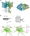
Figure 1
Figure 1
The chloride channel ClC-2. (A) The structural configuration of a CLC chloride channel. (Left) Lateral view of the dimeric form of the channel, which is comprised of two subunits distinguished by different colours. The intracellular cystathionine-β-synthase (CBS) domains are also highlighted in the unshaded box. (Right) Frontal view of the dimeric channel, where the two pores are highlighted in the purple boxes. Figures generated using the PDB model 5TQQ from human ClC-1, which has high homology to the ClC-2 channel. (B) Proposed model for depolarizing-mediated ClC-2 activation by MLC1/GlialCAM in astrocytes. In basal conditions, ClC-2 displays outwardly rectifying currents, promoting Cl efflux (left). Upon depolarization and the consequent entrance of K+ into the cell, the MLC1/GlialCAM complex binds to ClC-2, allowing GlialCAM to modify ClC-2-mediated currents, generating a Cl influx into the cell that may compensate the excess of K+ (right).
Figure 2
Figure 2
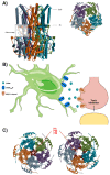
The bestrophin chloride channel. (A) The structural configuration of the Best1 channel. (Left) The schematic topology of Best1 channel, with the transmembrane and intracellular domains clearly identified and the calcium clasp highlighted in the box. (Right) Frontal view of the pentameric channel, where the five subunits are differentiated by colour and the central pore is visible in the centre of the channel. Figures generated using the PDB model 4RDQ from chicken Best1 protein. (B) Schematic model of Best1-mediated tonic inhibition in the cerebellum, where astrocytes release GABA through Best1, which acts on neuronal GABAergic receptors, which in turn may be relevant for motor coordination. (C) Closed (left) and open (right) configurations of the Best1 channel, with the residues conforming the hydrophobic seal (I76, F80, F84) highlighted in red. Note the significant change in the size of the pore in the open (Ca2+ bound) configuration. Figures were generated using the PDB models 6N23 and 6N28 from the structure of the chicken Best1 channel.
Figure 3
Figure 3
The VRAC. (A) The cryo-based model of the 8A-VRAC viewed from the membrane plane. The different subunits of the hexamer are highlighted in different colours, and domain layers are indicated by dashed rectangles. (B) Extracellular (top) and intracellular (bottom) views of the 8A-VRAC model. Dashed lines highlight the symmetry axis of the structures, with a six-fold symmetry axis observed in the top view, and a three-fold symmetry axis in the bottom view (LRRD). (C) Representation of a single LRRC8A subunit. Different helices and domains are indicated in different colours. Dashed squares delimit unresolved regions in the structure. Relevant positive residues (blue), negative (red) and polar (green) are indicated with coloured spheres. (D) The hypothetical model of the pathophysiological role of the astrocytic VRAC in ischaemia. Ischaemic conditions lead to depletion of intracellular ATP due to unsustainable oxidative metabolism, triggering the inactivation of the Na+/K+-pump, and subsequently cell swelling. NKCC1 cotransporter activity could further contribute to cell swelling. Astrocyte swelling triggers the opening of the LRRC8-VRAC mediating glutamate efflux. Glutamate build-up in the ischaemic penumbra produces its excitotoxic effect through the overactivation of glutamate-gated channels like NMDA, leading to sustained depolarization and increased Ca2+ levels, thus triggering neuronal damage and death. GPCR signalling could also modulate LRRC8-VRAC activation, but the possible contribution to the process is yet to be discovered. Pharmacological compounds like DCPIB could exert their protective effect by blocking the LRRC8-VRAC, thus reducing glutamate build-up in the ischaemic penumbra.
Figure 4
Figure 4
The SLCO2A1 protein, homology model and physiological role. (A) A 3D homology model of the SLCO2A1 protein based on the crystal structure of the E. coli glycerol-3-phosphate transporter (PDB ID: 1PW4A), constructed using the I-TASSER® software, as described elsewhere [139]. The protein is shown from the membrane plane. All the missense mutations found in pachydermoperiostosis patients are shown, including the charge-neutralized mutants, K613G and R560N. The thick dashed line indicates the central axis of the protein, and the thin dashed rectangle delimits a proximity threshold to the central axis. The residues that are close to (yellow), or far from (blue) the central axis, are highlighted with coloured circles. Notice the preferential location of most mutants to the vicinity of the central axis. (B) The top view of the homology model. The central cross represents the central axis, and the dashed circle delimits the proximity area. (C) The hypothetical physiological role of the SLCO2A1 protein. The horizontal dashed line in the centre of the cell highlights the dual behaviour of the protein either in the prostaglandin transporter (PGT) conformation (down) or the Maxi-Cl channel (MAC) conformation (up). Under resting conditions, the phosphorylated SLCO2A1 protein behaves as the PGT, allowing for PGE2 uptake and its clearance from the extracellular fluid. Various stimuli (osmotic swelling, hypoxia, salt stress, patch excision, etc.) can trigger protein dephosphorylation, and the activation of the SLCO2A1-MAC activity (up). Released ATP would be able to mediate autocrine or paracrine purinergic signalling.

Similar articles

Cited by

References

    1. Jentsch T.J., Stein V., Weinreich F., Zdebik A.A. Molecular structure and physiological function of chloride channels. Physiol. Rev. 2002;82:503–568. doi: 10.1152/physrev.00029.2001. - DOI - PubMed
    1. Nilius B., Droogmans G. Amazing chloride channels: an overview. Acta Physiol. Scand. 2003;177:119–147. doi: 10.1046/j.1365-201X.2003.01060.x. - DOI - PubMed
    1. Wilson C.S., Mongin A.A. The signaling role for chloride in the bidirectional communication between neurons and astrocytes. Neurosci. Lett. 2018;689:33–44. doi: 10.1016/j.neulet.2018.01.012. - DOI - PMC - PubMed
    1. Blaesse P., Airaksinen M.S., Rivera C., Kaila K. Cation-Chloride Cotransporters and Neuronal Function. Neuron. 2009;61:820–838. doi: 10.1016/j.neuron.2009.03.003. - DOI - PubMed
    1. Nedergaard M., Ransom B., Goldman S.A. New roles for astrocytes: Redefining the functional architecture of the brain. Trends Neurosci. 2003;26:523–530. doi: 10.1016/j.tins.2003.08.008. - DOI - PubMed

Substances