Review
. 2019 Feb 27;9(3):316.
doi: 10.3390/nano9030316.

Nanomaterials-Based Colorimetric Immunoassays

Affiliations
Review

Nanomaterials-Based Colorimetric Immunoassays

Lin Liu et al. Nanomaterials (Basel). .

Abstract

Colorimetric immunoassays for tumor marker detection have attracted considerable attention due to their simplicity and high efficiency. With the achievements of nanotechnology and nanoscience, nanomaterials-based colorimetric immunoassays have been demonstrated to be promising alternatives to conventional colorimetric enzyme-linked immunoassays. This review is focused on the progress in colorimetric immunoassays with the signal amplification of nanomaterials, including nanomaterials-based artificial enzymes to catalyze the chromogenic reactions, analyte-induced aggregation or size/morphology change of nanomaterials, nanomaterials as the carriers for loading enzyme labels, and chromogenic reactions induced by the constituent elements released from nanomaterials.

Keywords: colorimetric immunoassays; metal ions; nanomaterials; nanoparticle aggregation; nanozymes.


Conflict of interest statement

The authors declare no conflict of interest.

Figures

Figure 1
Figure 1
(A) Schematic illustration of the magnetic nanobead-based nano(e)zyme-linked immunosorbent assay (MagLISA). Reproduced with permission from [47]. Copyright American Chemical Society, 2018. (B) Schematic representation of the plasmonic nanosenor, in which the target molecule is anchored to the substrate by capture antibodies and recognized by other antibodies labeled with gold nanoclusters (AuNCs). Reproduced with permission from [50]. Copyright American Chemical Society, 2016.
Figure 1
Figure 1
(A) Schematic illustration of the magnetic nanobead-based nano(e)zyme-linked immunosorbent assay (MagLISA). Reproduced with permission from [47]. Copyright American Chemical Society, 2018. (B) Schematic representation of the plasmonic nanosenor, in which the target molecule is anchored to the substrate by capture antibodies and recognized by other antibodies labeled with gold nanoclusters (AuNCs). Reproduced with permission from [50]. Copyright American Chemical Society, 2016.
Figure 2
Figure 2
(A) Peroxidase-like activity of Pd-Ir cubes. Reproduced with permission from [54]. Copyright American Chemical Society, 2015. (B) Detection of prostate surface antigen (PSA) with Pd-Ir cubes-based enzyme-linked immunosorbent assay (Pd-Ir ELISA) and conventional horseradish peroxidase-based ELISA (HRP ELISA). Reproduced with permission from [54]. Copyright American Chemical Society, 2015. (C) Schematic illustration of the utilization of Pd−Ir NPs@GVs based ELISA for the detection of disease biomarkers. Reproduced with permission from [55]. Copyright American Chemical Society, 2017.
Figure 3
Figure 3
(A) Poly-(vinylpyrrolidone) (PVP)-capped Pt cubes for the colorimetric detection of Ag+ ions. Reproduced with permission from [58]. Copyright American Chemical Society, 2017. (B) Schematics showing (a) the fabrication of Au@Pt NPs in which Pt atoms are deposited onto an AuNP to form a conformal, thin Pt shell with thicknesses of 1–10 atomic layers and (b) two types of color signal generated from Au@PtnL NPs under different mechanisms. Reproduced with permission from [60]. Copyright American Chemical Society, 2017. (C) Scheme showing amplified lateral flow immunoassay (LFIA), where functionalized Pt nanocatalysts and biotinylated nanobody fragments are mixed with a plasma or serum sample. Reproduced with permission from [61]. Copyright American Chemical Society, 2018.
Figure 4
Figure 4
(A) The peroxidase-like activity of magnetic nanoparticles (MNPs) and two immunoassays based on the peroxidase activity of MNPs. Reproduced with permission from [62]. Copyright The Nature Publishing Group, 2007. (B) Magnetocontrolled enzyme-mediated reverse colorimetric immunosensing strategy. Reproduced with permission from [64]. Copyright American Chemical Society, 2013. (C) Immunoassay based on a nanocomposite entrapping both MNPs and Pt NPs in mesoporous carbon. Reproduced with permission from [65]. Copyright John Wiley and Sons, 2014.
Figure 5
Figure 5
Schematic showing the HRP/H2O2 and nanoceria mediated oxidation of ampliflu. (a) In the pH range of 4–7, HRP/H2O2 oxidizes ampliflu to a nonfluorescent final product (resazurin); (b) in contrast, nanoceria oxidizes ampliflu to the intermediate oxidation fluorescent product (resorufin) at pH 7; (c) while at or below pH 5.0, nanoceria yields the terminal oxidized nonfluorescent product, resazurin; (d,e) the ability of nanoceria to oxidize ampliflu to a stable fluorescent product in the pH range of 6–8 will facilitate its use in ELISA without the use of H2O2. Reproduced with permission from [70]. Copyright American Chemical Society, 2011.
Figure 6
Figure 6
(A) Schematic diagram for the immunomagnetic capture and colorimetric detection of V. parahaemolyticus. Reproduced with permission from [75]. Copyright Springer Nature, 2017. (B) The principles and procedures of the MnO2 NPs based immunosensor for α-fetoprotein (AFP) detection. (a) Schematic procedures for preparation of BSA-MnO2 NPs and Ab2-MnO2 NPs conjugate; (b) Schematic illustration for the tyramine signal amplification (TSA) system; (c) procedures for AFP detection through the MnO2 NPs-based immunosensor combined with magnetic separation. Reproduced with permission from [76]. Copyright Springer Nature, 2017. (C) Schematic illustration of the conventional chemical reaction for an unconventional application in the magnetically responsive colorimetric immunoassay using enzyme-responsive just-in-time generation of MnO2 nanocatalyst. Reproduced with permission from [77]. Copyright Springer Nature, 2018.
Figure 7
Figure 7
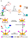
(A) Proposed immunodetection process for mouse IgG by coupling the cascade reaction of alkaline phosphatase (ALP) and the enzymatically in situ generated photoresponsive nanozyme of TiO2-CA. Reproduced with permission from [80]. Copyright American Chemical Society, 2017. (B) Proposed detection process using FA-CS-AgI. Reproduced with permission from [83]. Copyright American Chemical Society, 2017.
Figure 8
Figure 8
(A) Illustration of the colorimetric immunoassay of mIgG based on RIgG@Cu-MOF as a detection antibody. Reproduced with permission from [88]. Copyright American Chemical Society, 2018. (B) Illustration of the fabrication of FePor-TFPA-COP-based colorimetric immunoassay. Reproduced with permission from [93]. Copyright American Chemical Society, 2017.
Figure 9
Figure 9
(A) Naked-eye readout of plasmonic immunoassays. Detection of target protein via the combination of sandwich immunoassay, avidin−biotin interaction, glucose oxidase (GOx)-mediated oxidation of glucose, H2O2-induced oxidation of benzene-1,4-diboronic acid (BDBA), and BDBA-triggered aggregation of citrate-capped AuNPs. Reproduced with permission from [108]. Copyright American Chemical Society, 2016. (B) Schematic of the liposome-amplified plasmonic immunoassay. Reproduced with permission from [111]. Copyright American Chemical Society, 2015. (C) The AChE-catalyzed hydrolysis reaction for the colorimetric detection of enterovirus 71 (EV71). Reproduced with permission from [112]. Copyright John Wiley and Sons, 2013. (D) Plasmonic immunoassay based on HRP mediated modulation of AuNPs that enables naked-eye readout. Reproduced with permission from [114]. Copyright American Chemical Society, 2015.
Figure 9
Figure 9
(A) Naked-eye readout of plasmonic immunoassays. Detection of target protein via the combination of sandwich immunoassay, avidin−biotin interaction, glucose oxidase (GOx)-mediated oxidation of glucose, H2O2-induced oxidation of benzene-1,4-diboronic acid (BDBA), and BDBA-triggered aggregation of citrate-capped AuNPs. Reproduced with permission from [108]. Copyright American Chemical Society, 2016. (B) Schematic of the liposome-amplified plasmonic immunoassay. Reproduced with permission from [111]. Copyright American Chemical Society, 2015. (C) The AChE-catalyzed hydrolysis reaction for the colorimetric detection of enterovirus 71 (EV71). Reproduced with permission from [112]. Copyright John Wiley and Sons, 2013. (D) Plasmonic immunoassay based on HRP mediated modulation of AuNPs that enables naked-eye readout. Reproduced with permission from [114]. Copyright American Chemical Society, 2015.
Figure 10
Figure 10
Principle of peptide-ALP-AuNPs immunoassay for simultaneous detection of the inflammatory markers (IL-6, PCT, and CRP). Reproduced with permission from [119]. Copyright American Chemical Society, 2018.
Figure 11
Figure 11
(A) The detection of Cu2+ ions using click chemistry between two types of gold NPs, each modified with thiols terminated in an alkyne (1) or an azide (2) functional group. Reproduced with permission from [121]. Copyright John Wiley and Sons, 2008. (B) Copper-mediated amplification allows a readout of the immunoassay by the naked eye based on CuO-labeled antibody and click chemistry. Reproduced with permission from [122]. Copyright John Wiley and Sons, 2011. (C) Plasmonic nanosensor based on ALP-triggered CuAAC between azide- and alkyne-functionalized AuNPs and a naked-eye readout of plasmonic immunoassays based on ALP-triggered CuAAC through three-round amplification. Reproduced with permission from [123]. Copyright American Chemical Society, 2014.
Figure 12
Figure 12
(A) Schematic representation of the sandwich ELISA format used here and two possible signal generation mechanisms. Reproduced with permission from [34]. Copyright The Nature Publishing Group, 2012. (B) Schematic of the proposed quantitative immunoassay based on SiO2@PAA@CAT-catalyzed growth of AuNPs. Reproduced with permission from [139]. Copyright American Chemical Society, 2015. (C) Schematic diagram of the quantitative immunoassay based on glucose oxidase (GOx)-catalyzed growth of gold nanoparticles. Reproduced with permission from [31]. Copyright American Chemical Society, 2014. (D) Schematic representation of the sandwich plasmonic ELISA and the signal-generation method. Reproduced with permission from [126]. Copyright American Chemical Society, 2015. (E) Illustration of the protocol for the colorimetric magnetoimmunoassay of H9N2 AIV. Reproduced with permission from [128]. Copyright American Chemical Society, 2014.
Figure 13
Figure 13
(A) Schematic illustration of the high-resolution colorimetric assay for sensitive visual readout of phosphatase activity based on gold/silver core/shell nanorod. Reproduced with permission from [142]. Copyright American Chemical Society, 2014. (B) Schematic illustration for visual plasmonic ELISA based on ALP-triggered etching of AuNRs. Reproduced with permission from [144]. Copyright American Chemical Society, 2015. (C) Principle of the proposed naked-eye semiquantitative ELISA for visual quantification of proteins. Reproduced with permission from [147]. Copyright American Chemical Society, 2015.
Figure 14
Figure 14
(A) Schematic (not in scale) of the preparation of the complex Au-anti-CA15-3-HRP and the sandwich-type ELISA procedure without (IIIa) and with (IIIb) the application of AuNPs as the signal enhancer. Reproduced with permission from [153]. Copyright American Chemical Society, 2010. (B) Schematic illustration of serovar Typhimurium (STM) detection based on HRP and detection antibody-modified AuNPs. Reproduced with permission from [155]. Copyright American Chemical Society, 2014.
Figure 15
Figure 15
Scheme of the colorimetric strategy for the detection of ataxia telangiectasia mutated (ATM) using an MWNT-based probe compared with conventional ELISA. Reproduced with permission from [159]. Copyright American Chemical Society, 2011.
Figure 16
Figure 16
(A) Diagram of the drug delivery system inspired enzyme-linked immunosorbent assay (DDS-ELISA) for the detection of cTnI in 96-microwell plates. Reproduced with permission from [175]. Copyright American Chemical Society, 2018. (B) Synthesis and derivatization of TP@PEI/Ab2-MSNs and steps of the enzyme-free immunosorbent assay of PSA using TP@PEI/Ab2-MSNs for amplified colorimetric detection in a 96-well plate. Reproduced with permission from [176]. Copyright American Chemical Society, 2018. (C) Schematic representation of MPDA@TP-linked immunsorbent assay (MLISA) for α-fetoprotein (AFP) on anti-AFP capture antibody (CAb)-modified microplate by using anti-AFP detection antibody (DAb)-labeled MPDA@TP with a sandwich-type immunoreaction mode. Reproduced with permission from [177]. Copyright American Chemical Society, 2018.

References

    1. Farka Z., Jurik T., Kovar D., Trnkova L., Skladal P. Nanoparticle-based immunochemical biosensors and assays: Recent advances and challenges. Chem. Rev. 2017;117:9973–10042. doi: 10.1021/acs.chemrev.7b00037. - DOI - PubMed
    1. Li Z., Chen G.-Y. Current conjugation methods for immunosensors. Nanomaterials. 2018;8:278. doi: 10.3390/nano8050278. - DOI - PMC - PubMed
    1. Yin Y., Cao Y., Xu Y., Li G. Colorimetric immunoassay for detection of tumor markers. Int. J. Mol. Sci. 2010;11:5077–5094. doi: 10.3390/ijms11125077. - DOI - PMC - PubMed
    1. Zheng W., Jiang X. Integration of nanomaterials for colorimetric immunoassays with improved performance: A functional perspective. Analyst. 2016;141:1196–1208. doi: 10.1039/C5AN02222C. - DOI - PubMed
    1. Sun J., Xianyu Y., Jiang X. Point-of-care biochemical assays using gold nanoparticle-implemented microfluidics. Chem. Soc. Rev. 2014;43:6239–6253. doi: 10.1039/C4CS00125G. - DOI - PubMed

LinkOut - more resources