

Genetic Markers of ADHD-Related Variations in Intracranial Volume
- PMID: 30818988
- PMCID: PMC7780894
- DOI: 10.1176/appi.ajp.2018.18020149
Genetic Markers of ADHD-Related Variations in Intracranial Volume
Abstract
Objective: Attention deficit hyperactivity disorder (ADHD) is a common and highly heritable neurodevelopmental disorder with a complex pathophysiology. Intracranial volume (ICV) and volumes of the nucleus accumbens, amygdala, caudate nucleus, hippocampus, and putamen are smaller in people with ADHD compared with healthy individuals. The authors investigated the overlap between common genetic variation associated with ADHD risk and these brain volume measures to identify underlying biological processes contributing to the disorder.
Methods: The authors combined genome-wide association results from the largest available studies of ADHD (N=55,374) and brain volumes (N=11,221-24,704), using a set of complementary methods to investigate overlap at the level of global common variant genetic architecture and at the single variant level.
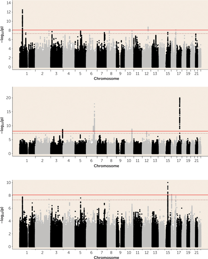
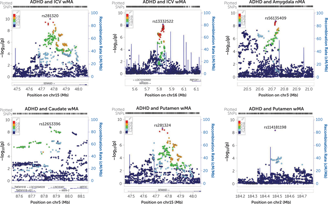
Results: Analyses revealed a significant negative genetic correlation between ADHD and ICV (rg=-0.22). Meta-analysis of single variants revealed two significant loci of interest associated with both ADHD risk and ICV; four additional loci were identified for ADHD and volumes of the amygdala, caudate nucleus, and putamen. Exploratory gene-based and gene-set analyses in the ADHD-ICV meta-analytic data showed association with variation in neurite outgrowth-related genes.
Conclusions: This is the first genome-wide study to show significant genetic overlap between brain volume measures and ADHD, both on the global and the single variant level. Variants linked to smaller ICV were associated with increased ADHD risk. These findings can help us develop new hypotheses about biological mechanisms by which brain structure alterations may be involved in ADHD disease etiology.
Keywords: ADHD; Genetics; MRI Brain Imaging.
Figures
Similar articles
-
Developmentally stable whole-brain volume reductions and developmentally sensitive caudate and putamen volume alterations in those with attention-deficit/hyperactivity disorder and their unaffected siblings.JAMA Psychiatry. 2015 May;72(5):490-9. doi: 10.1001/jamapsychiatry.2014.3162. JAMA Psychiatry. 2015. PMID: 25785435
-
Brain alterations in adult ADHD: effects of gender, treatment and comorbid depression.Eur Neuropsychopharmacol. 2014 Mar;24(3):397-409. doi: 10.1016/j.euroneuro.2013.11.011. Epub 2013 Dec 1. Eur Neuropsychopharmacol. 2014. PMID: 24345721
-
Subcortical brain volume differences in participants with attention deficit hyperactivity disorder in children and adults: a cross-sectional mega-analysis.Lancet Psychiatry. 2017 Apr;4(4):310-319. doi: 10.1016/S2215-0366(17)30049-4. Epub 2017 Feb 16. Lancet Psychiatry. 2017. PMID: 28219628 Free PMC article.
-
Integrated genome-wide association study findings: identification of a neurodevelopmental network for attention deficit hyperactivity disorder.Am J Psychiatry. 2011 Apr;168(4):365-77. doi: 10.1176/appi.ajp.2010.10070948. Epub 2011 Feb 15. Am J Psychiatry. 2011. PMID: 21324949 Review.
-
Meta-analysis of structural MRI studies in children and adults with attention deficit hyperactivity disorder indicates treatment effects.Acta Psychiatr Scand. 2012 Feb;125(2):114-26. doi: 10.1111/j.1600-0447.2011.01786.x. Epub 2011 Nov 28. Acta Psychiatr Scand. 2012. PMID: 22118249 Review.
Cited by
-
Mapping causal pathways from genetics to neuropsychiatric disorders using genome-wide imaging genetics: Current status and future directions.Psychiatry Clin Neurosci. 2019 Jul;73(7):357-369. doi: 10.1111/pcn.12839. Epub 2019 May 21. Psychiatry Clin Neurosci. 2019. PMID: 30864184 Free PMC article. Review.
-
Shared Genetic Determinants of Schizophrenia and Autism Spectrum Disorder Implicate Opposite Risk Patterns: A Genome-Wide Analysis of Common Variants.Schizophr Bull. 2024 Nov 8;50(6):1382-1395. doi: 10.1093/schbul/sbae044. Schizophr Bull. 2024. PMID: 38616054 Free PMC article.
-
ENIGMA and global neuroscience: A decade of large-scale studies of the brain in health and disease across more than 40 countries.Transl Psychiatry. 2020 Mar 20;10(1):100. doi: 10.1038/s41398-020-0705-1. Transl Psychiatry. 2020. PMID: 32198361 Free PMC article. Review.
-
Multivariate genome-wide analysis of education, socioeconomic status and brain phenome.Nat Hum Behav. 2021 Apr;5(4):482-496. doi: 10.1038/s41562-020-00980-y. Epub 2020 Dec 21. Nat Hum Behav. 2021. PMID: 33349686 Free PMC article.
-
Development of ADHD: Etiology, Heterogeneity, and Early Life Course.Annu Rev Dev Psychol. 2020 Dec;2(1):559-583. doi: 10.1146/annurev-devpsych-060320-093413. Epub 2020 Oct 23. Annu Rev Dev Psychol. 2020. PMID: 34368774 Free PMC article.
References
-
- Faraone SV, Asherson P, Banaschewski T, et al.: Attention-deficit/hyperactivity disorder. Nat Rev Dis Primers 2015; 1:15020. - PubMed
-
- Faraone SV, Perlis RH, Doyle AE, et al.: Molecular genetics of attention-deficit/hyperactivity disorder. Biol Psychiatry 2005; 57: 1313–1323 - PubMed
-
- Greven CU, Bralten J, Mennes M, et al.: Developmentally stable whole-brain volume reductions and developmentally sensitive caudate and putamen volume alterations in those with attention-deficit/hyperactivity disorder and their unaffected siblings. JAMA Psychiatry 2015; 72:490–499 - PubMed
Publication types
MeSH terms
Substances
Grants and funding
LinkOut - more resources
Full Text Sources
Medical
