Unexplained paraparesis following subarachnoid haemorrhage
- PMID: 30819681
- PMCID: PMC6398817
- DOI: 10.1136/bcr-2018-227666
Unexplained paraparesis following subarachnoid haemorrhage
Abstract
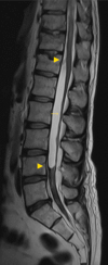
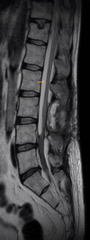
Spinal arachnoid cysts (SAC) are rare in isolation and the exact aetiology is still debated. Primary (congenital) cysts are caused by structural abnormalities in the arachnoid layer and largely affect the thoracic region. Secondary cysts are induced by a multitude of factors, infection, trauma or iatrogenic response, and can affect any level of the spinal cord. While subarachnoid haemorrhage (SAH) is a relatively common condition with significant repercussions, it is extremely uncommonly associated with SAC. When present, it may develop in the months and years after the original bleed, giving rise to new neurological symptoms. Prompt treatment is needed to halt or reverse the worsening of symptoms and questions are still being asked about how best to approach this condition. A 42-year-old man presented with chronic back pain, severe worsening ataxia and numbness below the umbilicus, 7 months after treatment for a World Federation of Neurosurgical Societies grade five (WFNS V) SAH. Imaging revealed a SAC extending from T12 to L4 and causing thecal compression. This was treated with a L3 laminectomy andmarsupialisation. An improvement in neurological function was observed at 6 months. Aetiology of the SAC and its association with SAH are discussed and a review of the relevant literature is provided.
Keywords: spinal cord; stroke.
© BMJ Publishing Group Limited 2019. No commercial re-use. See rights and permissions. Published by BMJ.
Conflict of interest statement
Competing interests: None declared.
Figures


References
Publication types
MeSH terms
LinkOut - more resources
Full Text Sources
Medical
