

doi: 10.3174/ajnr.A5995.
Epub 2019 Feb 28.
Decubitus CT Myelography for Detecting Subtle CSF Leaks in Spontaneous Intracranial Hypotension
Affiliations
- PMID: 30819772
- PMCID: PMC7048526
- DOI: 10.3174/ajnr.A5995
Item in Clipboard
Decubitus CT Myelography for Detecting Subtle CSF Leaks in Spontaneous Intracranial Hypotension
AJNR Am J Neuroradiol.
2019 Apr.
Abstract
Spontaneous intracranial hypotension is caused by spinal CSF leaks, but the site of the leak is not always detected on spinal imaging. We report on the additional value of decubitus positioning during CT myelography in enhancing the detection of subtle leaks.
© 2019 by American Journal of Neuroradiology.
Figures
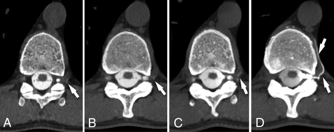
CSF venous fistula visualized best on decubitus CTM. Axial prone CTM image (A) and maximum-intensity-projection image of the same level (B) show subtle linear contrast (arrows) lateral to the T11 nerve root. The patient was turned to the left lateral decubitus position and re-scanned 13 minutes later. Axial MIP image (C) from that scan shows increased intravascular contrast with the patient in the decubitus position, suggestive of a CSF venous fistula (arrow). Axial MIP image (D) from a CTM performed after dynamic myelography on a subsequent day with the patient maintained in the decubitus position after contrast injection shows extensive filling of a paraspinal vein distal to the fistula (arrows).
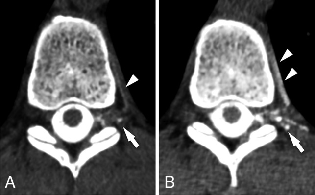
CSF venous fistula visualized best on decubitus CTM. Axial prone CT myelogram (A) shows subtle filling of a network of paraspinal veins (arrow) lateral to the nerve root and in the adjacent spinal segmental vein (arrowhead). Axial image (B) from a decubitus CTM performed after dynamic myelography on a subsequent day shows increased filling of the lateral veins (arrow) and segmental vein (arrowheads), making the CVF much more conspicuous.
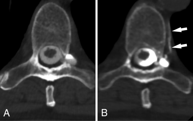
CSF venous fistula visualized best on decubitus CTM. Axial prone CTM image (A) shows possible increased density of a spinal segmental vein (arrow). Axial image (B) from a decubitus CTM performed after dynamic myelography on a subsequent day shows increased filling of the segmental veins (arrow), helping to confirm the diagnosis of CVF. Note the increased density of the left lateral thecal sac due to decubitus positioning.
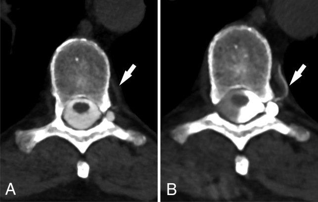
CSF venous fistula visualized best on decubitus CTM. Axial prone CTM image (A) shows a perineural diverticulum, but no clear leak. Axial image (B) from a subsequent CTM obtained with the patient in the decubitus position after dynamic myelography shows clear filling of a segmental vein (arrows), confirming a CVF.
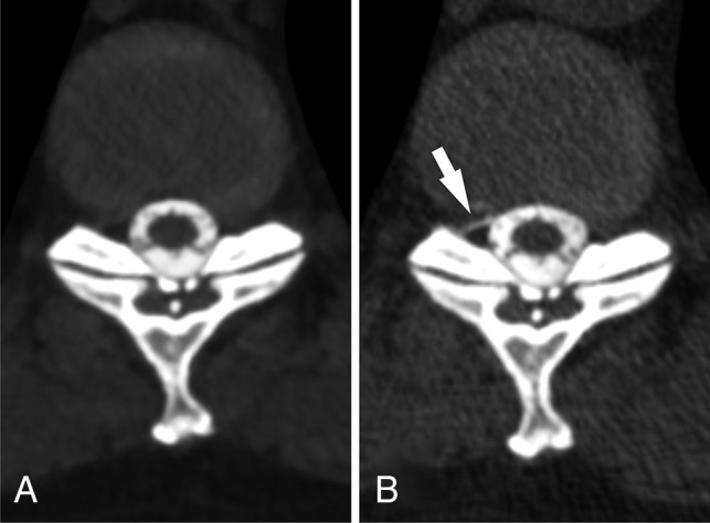
Low-flow CSF leak seen only on decubitus myelography. Axial prone CTM image (A) shows no CSF leak at the T10–11 level. The patient was turned into the left lateral decubitus position and re-scanned, again with the scan showing no leak (not shown). The patient was then turned to the right lateral decubitus position. An axial CTM image from the decubitus myelogram (B) obtained 6 minutes later shows a low-flow CSF leak not seen on prone myelogram (arrow).
References
MeSH terms
LinkOut - more resources
Full Text Sources