. 2019 Apr 19;294(16):6416-6429.
doi: 10.1074/jbc.RA118.006561. Epub 2019 Feb 28.

Molecular basis of cullin-3 (Cul3) ubiquitin ligase subversion by vaccinia virus protein A55

Affiliations

Molecular basis of cullin-3 (Cul3) ubiquitin ligase subversion by vaccinia virus protein A55

Chen Gao et al. J Biol Chem. .

Abstract

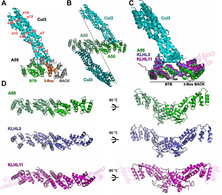
BTB-Kelch proteins are substrate-specific adaptors for cullin-3 (Cul3) RING-box-based E3 ubiquitin ligases, mediating protein ubiquitylation for subsequent proteasomal degradation. Vaccinia virus encodes three BTB-Kelch proteins: A55, C2, and F3. Viruses lacking A55 or C2 have altered cytopathic effects in cultured cells and altered pathology in vivo Previous studies have shown that the ectromelia virus orthologue of A55 interacts with Cul3 in cells. We report that the N-terminal BTB-BACK (BB) domain of A55 binds directly to the Cul3 N-terminal domain (Cul3-NTD), forming a 2:2 complex in solution. We solved the structure of an A55BB/Cul3-NTD complex from anisotropic crystals diffracting to 2.3/3.7 Å resolution in the best/worst direction, revealing that the overall interaction and binding interface closely resemble the structures of cellular BTB/Cul3-NTD complexes, despite low sequence identity between A55 and cellular BTB domains. Surprisingly, despite this structural similarity, the affinity of Cul3-NTD for A55BB was stronger than for cellular BTB proteins. Glutamate substitution of the A55 residue Ile-48, adjacent to the canonical φX(D/E) Cul3-binding motif, reduced affinity of A55BB for Cul3-NTD by at least 2 orders of magnitude. Moreover, Ile-48 and the φX(D/E) motif are conserved in A55 orthologues from other poxviruses, but not in the vaccinia virus proteins C2 or F3. The high-affinity interaction between A55BB and Cul3-NTD suggests that, in addition to directing the Cul3-RING E3 ligase complex to degrade cellular/viral target proteins that are normally unaffected, A55 may also sequester Cul3 from cellular adaptor proteins, thereby protecting substrates of these cellular adaptors from ubiquitylation and degradation.

Keywords: BTB–Kelch; E3 ubiquitin ligase; X-ray crystallography; immunosuppression; innate immunity; isothermal titration calorimetry (ITC); poxvirus; protein structure; structure-function; viral immunology.


Conflict of interest statement

The authors declare that they have no conflicts of interest with the contents of this article

Figures

Figure 1.
Figure 1.
A55 directly binds to cullin-3 via its N-terminal BB domain. A–C, representative immunoblots following immunoprecipitation (IP) of cleared lysates from HEK293T–REx cell lines (A) expressing empty vector (EV), B14–TAP (B14), or TAP–A55 (A55), the TAP tag comprising STREP and FLAG epitopes; (B) expressing B14–TAP or TAP–A55 and transfected with plasmids encoding myc-Cul3 or myc-Cul5; (C) expressing EV, B14–TAP, TAP–A55, TAP–A55BB, or TAP–A55 Kelch. Cells were lysed in Nonidet P-40 (A and C) or RIPA buffer (B). Immunoprecipitates were subjected to SDS-PAGE and immunoblotting. A and C, FLAG IP and immunoblotting for co-IP of endogenous Cul3. B, Myc IP and immunoblotting for co-IP of TAP-tagged B14 or A55. Input, cleared lysate. Data shown are representative of at least three independent experiments. Signals arising from the light chain (*) or heavy chain (**) of the antibody used for IP are marked. D, SEC-MALS analyses showing the SEC elution profiles (thin lines) and molecular mass distribution (thick lines) across the elution peaks for A55BB (peak 1, green, theoretical molecular mass 30 kDa and observed molecular mass 60 kDa), Cul3NΔ22 (peak 2, blue, theoretical molecular mass 46 kDa and observed molecular mass 45 kDa), and A55BB and Cul3NΔ22 together (peak 3, red, theoretical molecular mass 76 kDa and observed molecular mass 141 kDa) when eluting from a Superdex 200 10/300 GL column. Peak 4 is assumed to be excess Cul3NΔ22. E, Coomassie-stained SDS-PAGE analysis of peaks 1–3 from D.
Figure 2.
Figure 2.
ITC studies show that A55 binds to Cul3 with nanomolar to sub-nanomolar affinity. A–D, representative ITC titration curves showing interactions between A55BB and Cul3NΔ22 (A) or Cul3N (B) and between KLHL3, a human BTB related to A55, and Cul3NΔ22 (C) or Cul3N (D). The top figure in each panel shows the baseline-corrected differential power (DP) versus time. The bottom figure of each panel is the normalized binding curve showing integrated changes in enthalpy (ΔH) against molar ratio. The corresponding dissociation constant (KD), number of binding sites (N), enthalpy change (ΔH), and change in Gibbs free energy (ΔG) for each representative experiment are shown. All experiments were performed at least twice independently.
Figure 3.
Figure 3.
A55 and cellular BB domains share conserved modes of dimerization and Cul3 binding. A, structure of the A55/Cul3NΔ22 heterodimer in the asymmetric unit as ribbon diagram. Cul3 is in cyan and the three domains of A55 (BTB, three-box and BACK) are in green, orange, and gray, respectively. Helices α1–α12 from A55 are labeled in black with the exception of α10, which is hidden behind α9 in the picture. Helices α1–α16 from Cul3 are labeled in red. B, A55/Cul3 dimer formed by applying crystallographic 2-fold symmetry. C, overlay of three BB/Cul3 complex structures (KLHL3/Cul3, PDB code 4HXI (7); KLHL11/Cul3, PDB code 4APF (6); and A55/Cul3). The structures are aligned to the Cul3 part of the A55/Cul3 complex only. A55, KLHL3, KLHL11, and Cul3 are in green, purple, magenta, and cyan, respectively, and the three sub-domains are marked. Additional helices at the C terminus of the KLHL11 BACK domain are shown as semi-transparent helices. D, comparison of the dimers formed by A55, KLHL3, and KLHL11 BB domains, colored as in C.
Figure 4.
Figure 4.
Structure-based sequence alignment of the A55, KLHL3, and KLHL11 BB domains. Columns are shaded based on amino acid similarity. Secondary structural elements for A55 are shown above the aligned sequences and colored as in Fig. 3A. Residues at the A55/Cul3 interface are underlined in blue. Residues selected for subsequent mutagenesis studies of A55 are marked by stars at bottom: the two conserved sites (Phe-54 and Asp-56) are marked by purple stars, and the nonconserved site (Ile-48) is marked by a red star. Residues from KLHL11 that are involved in Cul3–NTE binding are marked by magenta triangles.
Figure 5.
Figure 5.
Conserved and nonconserved interactions at the interface between A55 and Cul3. A, A55BB/Cul3NΔ22 complex structure with two key Cul3-binding sites in the BTB domain boxed in black (enlarged in B–F) and red (enlarged in G). B, structural overlay of the φX(D/E) motifs from A55, KLHL3, KLHL11, and SPOP. C–F, surface of Cul3 colored by residue hydrophobicity from yellow (hydrophobic) to white (polar) (70). Hydrophobic binding pockets are shown for Phe-54 of A55, Met-83 of KLHL3, Phe-130 of KLHL11, and Met-233 of SPOP, which are equivalent to the φ residue of the φX(D/E) motif, and for Ile-48 of A55 and its equivalent residues Ala-77, Pro-124, and Ala-227 in KLHL3, KLHL11, and SPOP, respectively. G, overlay of the hydrogen bond formed between Tyr-125 of Cul3 and Asp-99 of A55 with equivalent residues Ser-128, Asp-181, and Asp-278 in KLHL3, KLHL11, and SPOP, respectively.
Figure 6.
Figure 6.
I48E mutation significantly impairs A55 binding to Cul3. A, representative thermal melt curves of WT A55BB and mutants F54E, D99A, I48A, and I48E from DSF studies. Curves are offset along the vertical axis for clarity. All experiments were performed in triplicate. B, comparison of the melting temperatures for WT A55BB (green), F54E (red), D99A (blue), I48A (orange), and I48E (black) mutants. Upper and lower panels display Tm values for the first and second melting events, respectively. Error bars show the standard errors of the mean from experiments performed in triplicate. C–J, representative ITC titration curves showing binding of A55BB mutants F54E (C and D), D99A (E and F), I48A (G and H), and I48E (I and J) to Cul3NΔ22 and Cul3N, respectively. Integrated changes in enthalpy (ΔH) are plotted against molar ratio of titrant. The corresponding dissociation constant (KD), number of binding sites (N), enthalpy change (ΔH), and change in Gibbs free energy (ΔG) for each representative experiment are shown. All experiments were performed at least twice independently. Raw data for C–J are shown in Fig. S7.
Figure 7.
Figure 7.
Cul3-binding residues of A55 are conserved across orthopoxvirus orthologues but not in VACV paralogues C2 and F3. Multiple sequence alignment of the A55 BB domains against its orthologues from selected poxviruses and two other VACV BTB–Kelch proteins, C2 and F3. Columns are shaded based on amino acid similarity. Secondary structural elements for A55 are shown above the aligned sequences and colored as in Fig. 3A. Residues at the A55/Cul3 interface are underlined in blue. Residues aligned with A55-Ile-48 and the φX(D/E) motif are boxed in red.

References

    1. Fenner F., Anderson D. A., Arita I., Jezek Z., and Ladnyi I. D. (1988) Smallpox and its eradication. World Health Organization, Geneva, Switzerland
    1. Smith G. L., Benfield C. T., Maluquer de Motes C., Mazzon M., Ember S. W., Ferguson B. J., and Sumner R. P. (2013) Vaccinia virus immune evasion: mechanisms, virulence and immunogenicity. J. Gen. Virol. 94, 2367–2392 10.1099/vir.0.055921-0 - DOI - PubMed
    1. Bahar M. W., Graham S. C., Chen R. A., Cooray S., Smith G. L., Stuart D. I., and Grimes J. M. (2011) How vaccinia virus has evolved to subvert the host immune response. J. Struct. Biol. 175, 127–134 10.1016/j.jsb.2011.03.010 - DOI - PMC - PubMed
    1. Beard P. M., Froggatt G. C., and Smith G. L. (2006) Vaccinia virus kelch protein A55 is a 64-kDa intracellular factor that affects virus-induced cytopathic effect and the outcome of infection in a murine intradermal model. J. Gen. Virol. 87, 1521–1529 10.1099/vir.0.81854-0 - DOI - PubMed
    1. Pintard L., Willems A., and Peter M. (2004) Cullin-based ubiquitin ligases: Cul3–BTB complexes join the family. EMBO J. 23, 1681–1687 10.1038/sj.emboj.7600186 - DOI - PMC - PubMed

Publication types

MeSH terms