. 2019 Apr 1;33(7-8):466-476.
doi: 10.1101/gad.316554.118. Epub 2019 Feb 28.

Auxin regulates endosperm cellularization in Arabidopsis

Affiliations

Auxin regulates endosperm cellularization in Arabidopsis

Rita A Batista et al. Genes Dev. .

Abstract

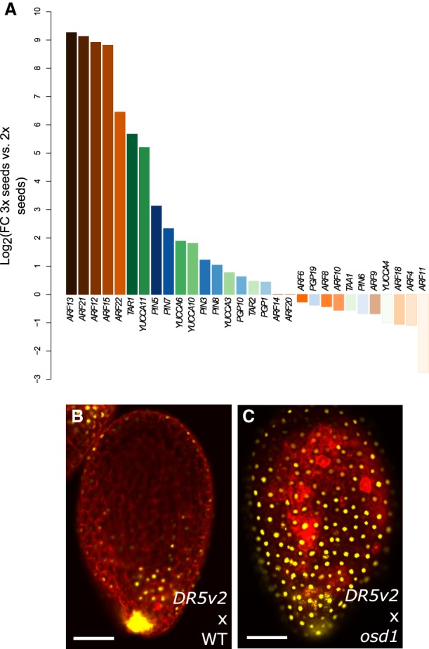
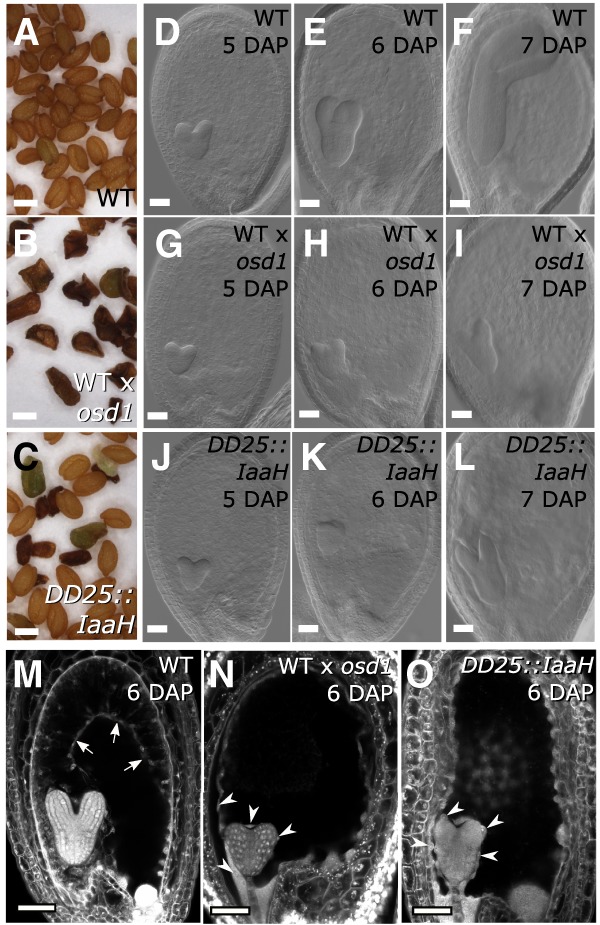
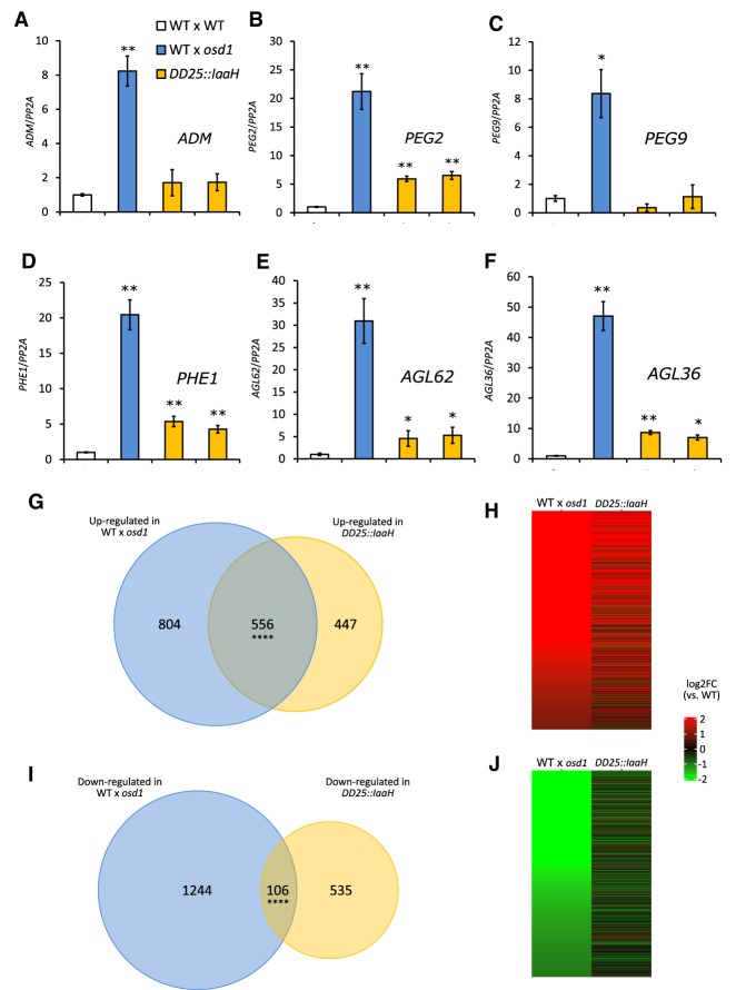
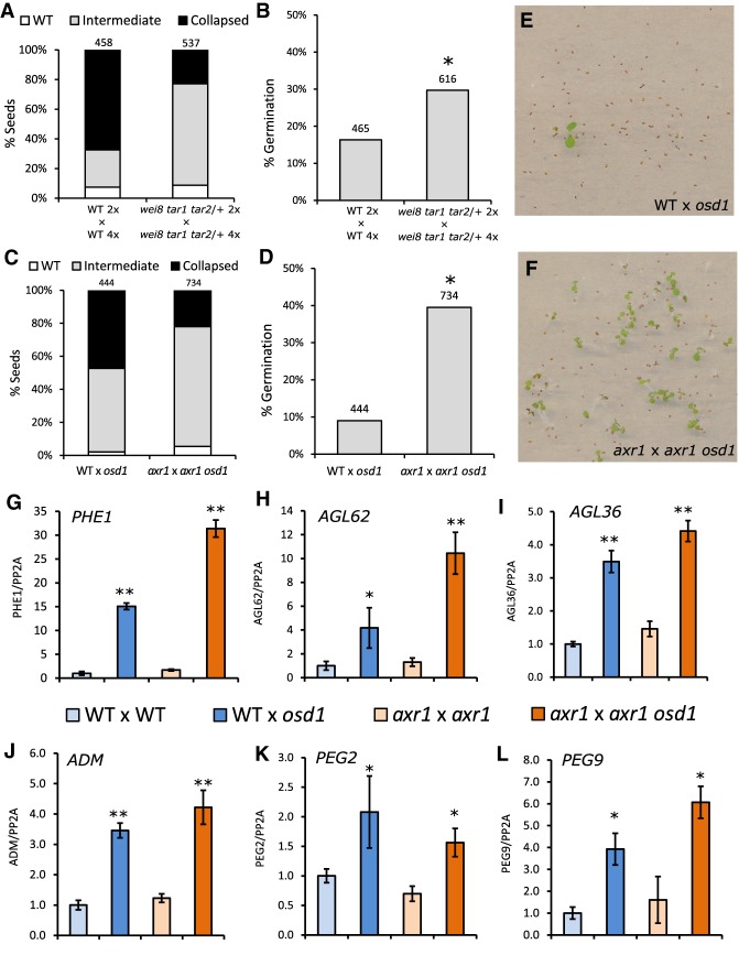
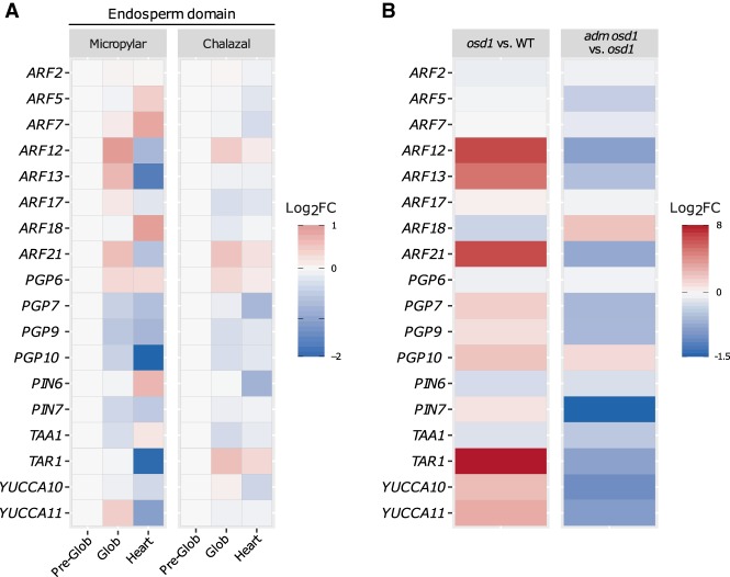
The endosperm is an ephemeral tissue that nourishes the developing embryo, similar to the placenta in mammals. In most angiosperms, endosperm development starts as a syncytium, in which nuclear divisions are not followed by cytokinesis. The timing of endosperm cellularization largely varies between species, and the event triggering this transition remains unknown. Here we show that increased auxin biosynthesis in the endosperm prevents its cellularization, leading to seed arrest. Auxin-overproducing seeds phenocopy paternal-excess triploid seeds derived from hybridizations of diploid maternal plants with tetraploid fathers. Concurrently, auxin-related genes are strongly overexpressed in triploid seeds, correlating with increased auxin activity. Reducing auxin biosynthesis and signaling reestablishes endosperm cellularization in triploid seeds and restores their viability, highlighting a causal role of increased auxin in preventing endosperm cellularization. We propose that auxin determines the time of endosperm cellularization, and thereby uncovered a central role of auxin in establishing hybridization barriers in plants.

Keywords: auxin; cellularization; endosperm; hybridization barrier; seed development; triploid block.


Figures

Figure 1.
Figure 1.
Auxin is overproduced in 3x seeds. (A) Log2-fold expression change between 3x and 2x seeds of genes coding for auxin biosynthesis (green bars), signaling (orange bars), and transport proteins (blue bars). (B,C) Auxin activity as measured by expression of DR5v2::VENUS in 2x (B) and 3x (C) seeds at 5 DAP. Pictures show representative seeds of three independent siliques per cross. Red staining is propidium iodide. Bars, 100 µm.
Figure 2.
Figure 2.
Increased auxin in the endosperm prevents cellularization. (AC) Dry seed morphology of wild-type 2x (A), wild-type 3x (B), and DD25::IaaH 2x seeds (C). Bars, 0.5 mm. (DL) Clearings of wild-type 2x (DF), wild-type 3x (GI), and DD25::IaaH 2x seeds (JL) from 5 to 7 DAP. Pictures show representative seeds of three independent siliques per cross. Bars, 50 µm. (MO) Endosperm cellularization as determined by Feulgen staining at 6 DAP for 2x seeds (M), 3x seeds (N), and 2x seeds expressing DD25::IaaH (O). Pictures show representative seeds of 10 independent siliques per cross. Arrows indicate cellularized peripheral endosperm, and arrowheads indicate free endosperm nuclei surrounding the embryo. Bars, 50 µm. (WT) Wild type.
Figure 3.
Figure 3.
PEGs and AGL genes are not substantially deregulated in DD25::IaaH transgenic seeds. (AF) Relative gene expression in seeds at 6 DAP, as determined by RT-qPCR, in 2x wild-type, 3x wild-type, and 2x DD25::IaaH transgenic seeds of two independent lines for ADM (A), PEG2 (B), PEG9 (C), PHE1 (D), AGL62 (E), and AGL36 (F). Results from a representative biological replicate are shown. Three technical replicates were performed; error bars, SD. Differences are significant for Student's t-test for P < 0.05 (*) or P < 0.001 (**). (WT) Wild-type. (GJ) Comparison of genes deregulated at 6 DAP, as determined by RNAseq, in 3x (WT × osd1) and 2x DD25::IaaH seeds. (G,I) The Venn diagrams show the overlap of genes that are commonly up-regulated (G) or down-regulated (I) in 3x and in 2x DD25::IaaH seeds. Overlaps between deregulated genes are significantly higher than expected; hypergeometric distribution, P < 0.0001 (****). (H,J) Each line in each plot indicates one gene that is deregulated in 3x seeds (left) and its corresponding expression in 2x seeds expressing DD25::IaaH (right).
Figure 4.
Figure 4.
Mutants in auxin biosynthesis and signaling suppress triploid seed abortion. (A,B) Phenotypic classification of 3x seeds in wild-type background and in the wei8 tar1 tar2 auxin biosynthesis mutant (A) and their corresponding germination rate (B). (C,D) Same as for A and B, but for the auxin-signaling-mutant axr1. Seed classification was performed according to that in Supplemental Figure S4. Numbers at the top indicate the number of seeds assayed. Differences between wild-type and mutant seed germination in B and D are significant for χ2 test for P < 0.0001 (*). (E,F) Representative image of germinating triploid seedlings in wild-type (E) and axr1 (F). (GL) Relative gene expression in 6-DAP seeds, as determined by RT-qPCR, in 2x and 3x seeds in wild-type and axr1 mutant backgrounds, for PHE1 (G), AGL62 (H), AGL36 (I), ADM (J), PEG2 (K), and PEG9 (L). Results of a representative biological replicate are shown. Three technical replicates were performed; error bars, SD. Differences between 3x seeds and each respective 2x control are significant for Student's t-test for P < 0.05 (*) or P < 0.001(**).
Figure 5.
Figure 5.
Endosperm cellularization is associated with down-regulation of auxin-related gene expression. (A) Expression of auxin-related genes in the micropylar and chalazal endosperm domains throughout different stages of seed development. Expression in each domain is normalized to the preglobular stage and expressed as a log2-fold change relative to that stage. (B) Log2-fold change of expression of auxin-related genes in 3x (osd1) versus 2x seeds and in 3x adm seeds (osd1 adm) versus 3x seeds (osd1).

Similar articles

Cited by

References

    1. Adamski NM, Anastasiou E, Eriksson S, O'Neill CM, Lenhard M. 2009. Local maternal control of seed size by KLUH/CYP78A5-dependent growth signaling. Proc Natl Acad Sci 106: 20115–20120. 10.1073/pnas.0907024106 - DOI - PMC - PubMed
    1. Anders S, Huber W. 2010. Differential expression analysis for sequence count data. Genome Biol 11: R106 10.1186/gb-2010-11-10-r106 - DOI - PMC - PubMed
    1. Belmonte MF, Kirkbride RC, Stone SL, Pelletier JM, Bui AQ, Yeung EC, Hashimoto M, Fei J, Harada CM, Munoz MD, et al. 2013. Comprehensive developmental profiles of gene activity in regions and subregions of the Arabidopsis seed. Proc Natl Acad Sci 110: E435–E444. 10.1073/pnas.1222061110 - DOI - PMC - PubMed
    1. Bleckmann A, Alter S, Dresselhaus T. 2014. The beginning of a seed: regulatory mechanisms of double fertilization. Front Plant Sci 5: 452 10.3389/fpls.2014.00452 - DOI - PMC - PubMed
    1. Boer DR, Freire-Rios A, van den Berg WAM, Saaki T, Manfield IW, Kepinski S, López-Vidrieo I, Franco-Zorrilla JM, de Vries SC, Solano R, et al. 2014. Structural basis for DNA binding specificity by the auxin-dependent ARF transcription factors. Cell 156: 577–589. 10.1016/j.cell.2013.12.027 - DOI - PubMed