. 2019 Mar 19;116(12):5493-5498.
doi: 10.1073/pnas.1811929116. Epub 2019 Feb 28.

Cryo-electron microscopy structure of the filamentous bacteriophage IKe

Affiliations

Cryo-electron microscopy structure of the filamentous bacteriophage IKe

Jingwei Xu et al. Proc Natl Acad Sci U S A. .

Abstract

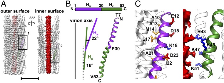
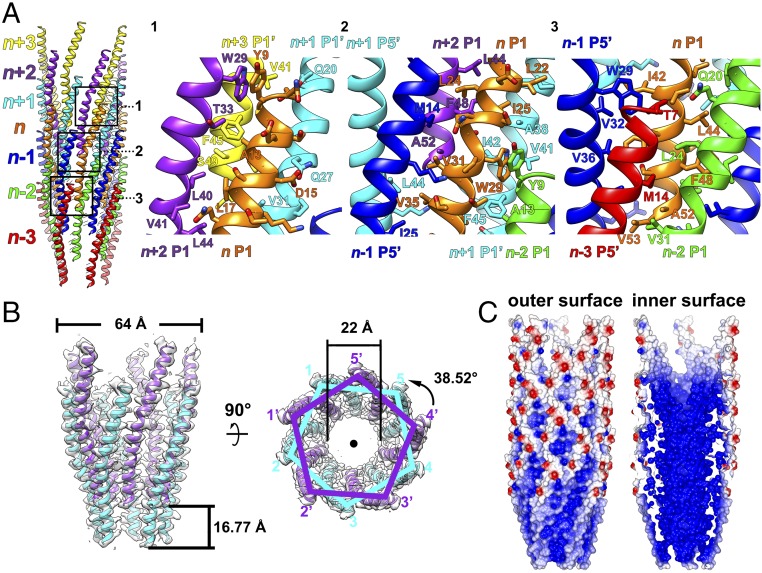
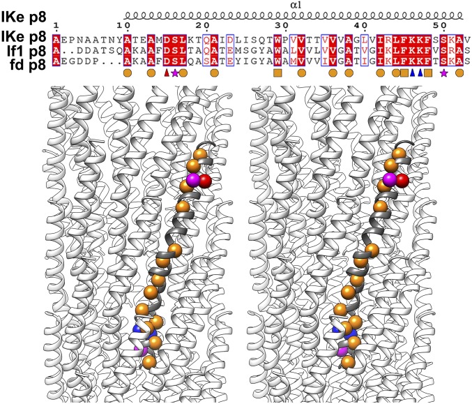
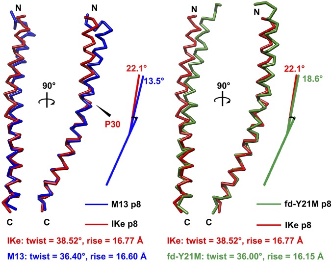
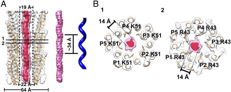
The filamentous bacteriophage IKe infects Escherichia coli cells bearing IncN pili. We report the cryo-electron microscopy structure of the micrometer-long IKe viral particle at a resolution of 3.4 Å. The major coat protein [protein 8 (p8)] consists of 47 residues that fold into a ∼68-Å-long helix. An atomic model of the coat protein was built. Five p8 helices in a horizontal layer form a pentamer, and symmetrically neighboring p8 layers form a right-handed helical cylinder having a rise per pentamer of 16.77 Å and a twist of 38.52°. The inner surface of the capsid cylinder is positively charged and has direct interactions with the encapsulated circular single-stranded DNA genome, which has an electron density consistent with an unusual left-handed helix structure. Similar to capsid structures of other filamentous viruses, strong capsid packing in the IKe particle is maintained by hydrophobic residues. Despite having a different length and large sequence differences from other filamentous phages, π-π interactions were found between Tyr9 of one p8 and Trp29 of a neighboring p8 in IKe that are similar to interactions observed in phage M13, suggesting that, despite sequence divergence, overall structural features are maintained.

Keywords: cryo-EM structure; filamentous virus; helical reconstruction; single-stranded circular DNA; symmetry mismatch.


Conflict of interest statement

The authors declare no conflict of interest.

Figures

Fig. 1.
Fig. 1.
Structure of the filamentous phage IKe. (A) Ribbon and surface shadowed diagrams showing the cryo-EM structure of the filamentous phage IKe. The helical capsid shell is semitransparent and colored white. The inner smeared DNA core is colored red. (Scale bar, 5 nm.) (B, Top) A schematic diagram showing the polypeptide chain of the IKe major coat protein p8. (B, Bottom) Ribbon diagrams showing the backbone of a single p8 monomer structure. The HN and HC of p8 are colored purple and green, respectively. The angle between HC and the virion axis and the hinge angle between HN and HC were measured, and values are shown in the schematic diagrams. (C) Ribbon and surface shadowed diagrams showing the hydrophobic and hydrophilic residues of HN (Left, position 1 on A) and the four positively charged residues of HC (Right, position 2 on A). The side chains of the hydrophobic, positively charged, and negatively charged residues are shown and colored orange, blue, and red, respectively. Close interactions with the DNA of positively charged R43 and K51 are apparent from the image.
Fig. 2.
Fig. 2.
Helical assembly of the IKe capsid. (A, Left) Ribbon diagram showing an IKe capsid fragment of seven pentameric protein layers (n + 3, n + 2, n + 1, n, n − 1, n − 2, n − 3) in different colors. (A, Right) Ribbon and stick diagrams of numbered boxed areas in the Left panel showing the interactions of one p8 monomer (n P1, in orange) with neighboring subunits. The major coat proteins from the same pentameric protein layer are colored identically. (B) Ribbon and surface shadowed diagrams showing side (Left) and top (Right) views of two pentameric protein layers and their relationship via helical symmetry operators. (C) Surface electrostatic potential of the p8 helical shell. Negative and positive electrostatic potentials are colored red and blue, respectively.
Fig. 3.
Fig. 3.
Sequence alignment of three filamentous bacteriophage major coat proteins. (Top) Sequence alignments of the major coat proteins of IKe, If1, and fd. The secondary structure is shown above the alignments. Completely conserved residues are shown in white on a red background. Conserved residues are boxed. Completely conserved hydrophobic residues with short side chains, aromatic residues, positively charged residues, negatively charged residues, and polar residues are marked by orange circles, orange cubes, blue triangles, red triangles, and magenta stars, respectively. (Bottom) Stereo ribbon diagrams showing the locations of the completely conserved residues in the IKe p8 structure. The conserved residues are represented by balls that are colored as described in the legend for the Top panel.
Fig. 4.
Fig. 4.
Structural comparisons of the major coat proteins of class I phages IKe, M13, and the fd-Y21M mutant. (Left) Structural superimpositions of the IKe major coat protein (red) with that of M13 (PDB entry 2MJZ, blue) based on the HC regions. (Right) Structure superimpositions of the IKe major coat protein (red) with that of phage fd Y21M mutant (PDB entry 2C0X, green) based on the HC regions. The helical parameters are given for each phage structure.
Fig. 5.
Fig. 5.
Structure of the inner ssDNA core. (A, Left) Ribbon and surface shadowed diagram showing the asymmetric reconstruction of the filamentous bacteriophage IKe calculated from the original raw images with the orientations determined for the inner DNA core. The helical shell is colored white, and the DNA core density contoured at a level of 17 σ is transparent and colored pink. The DNA core density contoured at a level of 26 σ is colored red. The model of the capsid is fitted in the map. (Middle and Right) Surface shadowed diagram showing the helical features of the inner DNA core and the left-handed helix. The left-handed helical pitch of the inner DNA core is ∼34 Å. (B) Ribbon and surface shadowed diagrams showing thin sections of the virus. The interactions between the capsid shell and the inner DNA core are mainly mediated through residues Lys51 and Arg43. The Arg43 residues are likely in contact with the DNA backbone. The densities from the shells are transparent and colored as described in A. The structure of IKe capsid shell is colored orange.

References

    1. Marzec CJ, Day LA. DNA and protein lattice-lattice interactions in the filamentous bacteriophages. Biophys J. 1983;42:171–180. - PMC - PubMed
    1. Day LA, Marzec CJ, Reisberg SA, Casadevall A. DNA packing in filamentous bacteriophages. Annu Rev Biophys Biophys Chem. 1988;17:509–539. - PubMed
    1. Rakonjac J, Bennett NJ, Spagnuolo J, Gagic D, Russel M. Filamentous bacteriophage: Biology, phage display and nanotechnology applications. Curr Issues Mol Biol. 2011;13:51–76. - PubMed
    1. Marvin DA. X-ray diffraction and electron microscope studies on the structure of the small filamentous bacteriophage fd. J Mol Biol. 1966;15:8–17. - PubMed
    1. Marzec CJ, Day LA. A theory of the symmetries of filamentous bacteriophages. Biophys J. 1988;53:425–440. - PMC - PubMed

Publication types

MeSH terms

Associated data