. 2019 Mar 19;116(12):5633-5642.
doi: 10.1073/pnas.1820320116. Epub 2019 Feb 28.

Regulatory changes in pterin and carotenoid genes underlie balanced color polymorphisms in the wall lizard

Affiliations

Regulatory changes in pterin and carotenoid genes underlie balanced color polymorphisms in the wall lizard

Pedro Andrade et al. Proc Natl Acad Sci U S A. .

Abstract

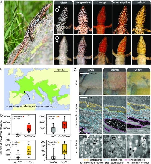
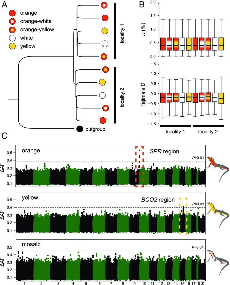
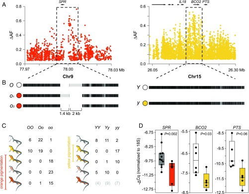
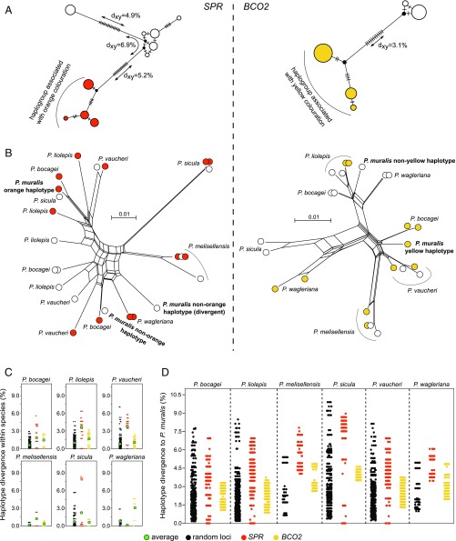
Reptiles use pterin and carotenoid pigments to produce yellow, orange, and red colors. These conspicuous colors serve a diversity of signaling functions, but their molecular basis remains unresolved. Here, we show that the genomes of sympatric color morphs of the European common wall lizard (Podarcis muralis), which differ in orange and yellow pigmentation and in their ecology and behavior, are virtually undifferentiated. Genetic differences are restricted to two small regulatory regions near genes associated with pterin [sepiapterin reductase (SPR)] and carotenoid [beta-carotene oxygenase 2 (BCO2)] metabolism, demonstrating that a core gene in the housekeeping pathway of pterin biosynthesis has been coopted for bright coloration in reptiles and indicating that these loci exert pleiotropic effects on other aspects of physiology. Pigmentation differences are explained by extremely divergent alleles, and haplotype analysis revealed abundant transspecific allele sharing with other lacertids exhibiting color polymorphisms. The evolution of these conspicuous color ornaments is the result of ancient genetic variation and cross-species hybridization.

Keywords: Podarcis muralis; balanced polymorphism; carotenoid pigmentation; introgression; pterin pigmentation.


Conflict of interest statement

The authors declare no conflict of interest.

Figures

Fig. 1.
Fig. 1.
Color polymorphism in the European common wall lizard, P. muralis. (A, Left) Common wall lizard. (A, Right) Illustrations of the five discrete ventral morphs. These conspicuous colors likely function as visual ornaments implicated in sexual signaling. The yellow and orange colors are restricted to the ventral surface, and males and females exhibit marked differences in the extent of pigmentation in some populations. (B) Geographic distribution of the species in Europe (light green). (C) Ultrastructure of the ventral skin of the three pure morphs. (Top) Close-up view of the ventral scales of each morph under a light microscope. (Middle) TEM of the three chromatophore layers [xantophores (yellow), iridophores (blue), and melanophores (pink)]. (Bottom) Electromicrographs detailing the structure of xanthophores. Examples of pterinosomes (pte), carotenoid vesicles (cv), and immature vesicles (im) are highlighted. (D) Levels of colored pterin and carotenoid compounds in the ventral skin of the different morphs obtained by HPLC-MS/MS. O, orange; W, white; Y, yellow.
Fig. 2.
Fig. 2.
Population structure and genetic basis of color polymorphism in the common wall lizard. (A) Neighbor-joining tree summarizing genetic distance among individual pools using 250,000 randomly chosen SNPs. The tree was rooted with a DNA pool of individuals sampled from Italy and belonging to a different intraspecific sublineage. (B) Nucleotide diversity (π) and Tajima’s D estimated for each morph. Both statistics were calculated in 10-kb nonoverlapping windows, and a genome-wide estimate was obtained by averaging all windows across the genome. (C) Genetic mapping based on differences in allele frequencies (ΔAF) for the orange, yellow, and mosaic phenotypes. The Manhattan plots show the median value of 20-SNP windows (five-SNP overlap) across the reference genome. The dashed lines represent a 1% significance cutoff based on 1,000 permutations conducted for each dataset.
Fig. 3.
Fig. 3.
Regulatory variation explains color polymorphism in the common wall lizard. (A) Differences in allele frequencies (ΔAF) for the orange and yellow phenotypes around SPR and BCO2 (each dot represents an SNP). (B) Haplotype structure for the same two regions based on the alignment of our reference genome sequence to consensus sequences of the alternative haplotypes obtained using Nanopore and Sanger sequencing. Black indicates homology, and light gray indicates mismatches that can originate from point mutations or indel variants. (C) Individual genotypes for the SPR and BCO2 loci among the five morphs based on high ΔAF variants selected from the whole-genome data. (D) qPCR measurements of SPR, BCO2, and PTS expression in ventral skin.
Fig. 4.
Fig. 4.
Evolution of the regulatory regions associated with pterin- and carotenoid-based coloration in the genus Podarcis. (A) Median-joining genealogies of the genomic regions associated with coloration upstream of SPR (Left) and BCO2 (Right) from common wall lizards from the Pyrenees. The dashes on branches indicate the number of observed mutations. (B) NeighborNet trees of the genomic regions associated with coloration upstream of SPR (Left) and BCO2 (Right) combining common wall lizards from the Pyrenees and six other species in the genus Podarcis. Haplotypes are colored orange, yellow, or white, indicating the color morph of the individual. For representation purposes, only a subset of the sequences is presented. (C) Pairwise nucleotide differences between haplotypes within species for SPR (orange), BCO2 (yellow), and 31 random loci (black). The average is indicated by a green circle. (D) Pairwise nucleotide differences between haplotypes of the common wall lizard and other Podarcis species for SPR (orange), BCO2 (yellow), and 31 random loci (black). The contrasts involving other species are presented in SI Appendix, Fig. S7.

Similar articles

Cited by

References

    1. McKinnon JS, Pierotti MER. Colour polymorphism and correlated characters: Genetic mechanisms and evolution. Mol Ecol. 2010;19:5101–5125. - PubMed
    1. Wellenreuther M, Svensson EI, Hansson B. Sexual selection and genetic colour polymorphisms in animals. Mol Ecol. 2014;23:5398–5414. - PubMed
    1. Sinervo B, Lively CM. The rock-paper-scissors game and the evolution of alternative male strategies. Nature. 1996;380:240–243.
    1. Sinervo B, Svensson E. Correlational selection and the evolution of genomic architecture. Heredity (Edinb) 2002;89:329–338. - PubMed
    1. Roulin A. The evolution, maintenance and adaptive function of genetic colour polymorphism in birds. Biol Rev Camb Philos Soc. 2004;79:815–848. - PubMed

Publication types

LinkOut - more resources