. 2019 Mar 19;116(12):5570-5575.
doi: 10.1073/pnas.1814930116. Epub 2019 Feb 28.

Neural crest-derived neurons invade the ovary but not the testis during mouse gonad development

Affiliations

Neural crest-derived neurons invade the ovary but not the testis during mouse gonad development

Jennifer McKey et al. Proc Natl Acad Sci U S A. .

Abstract

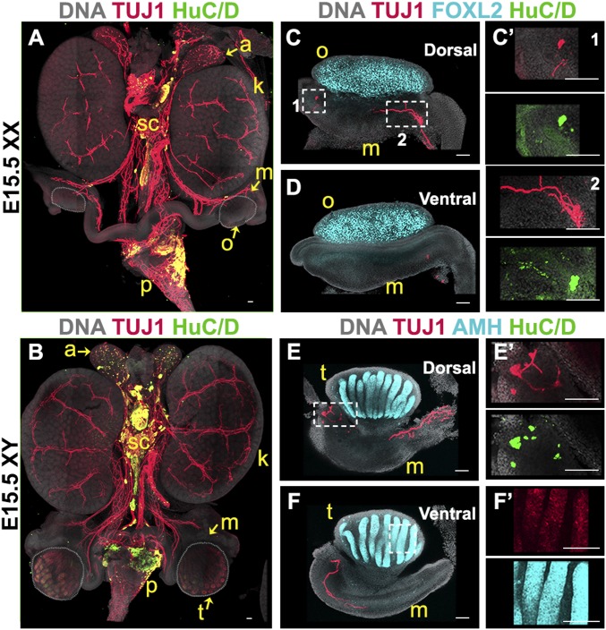
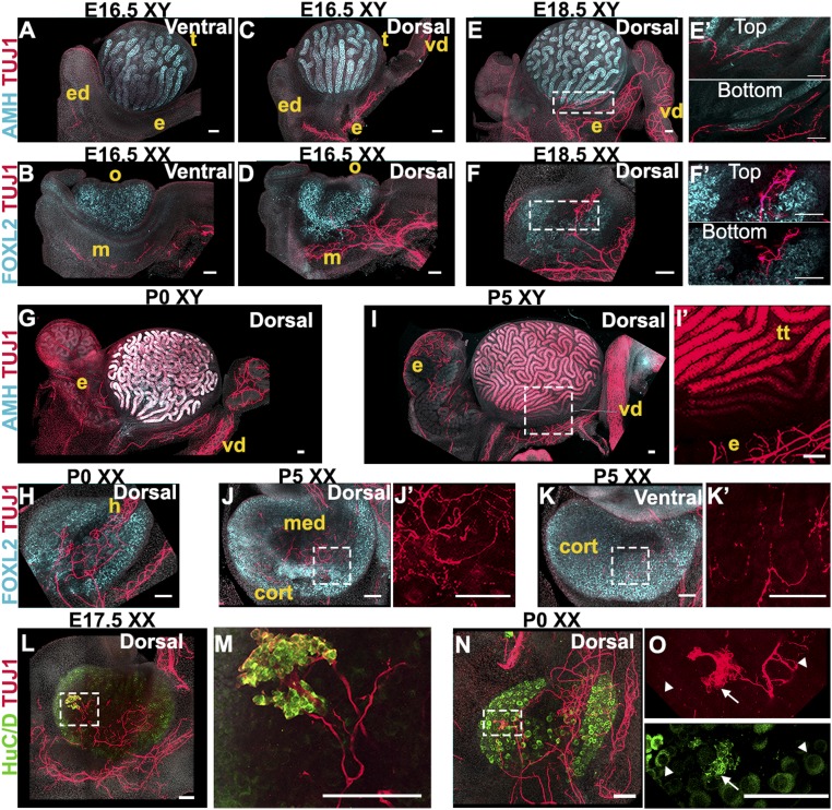
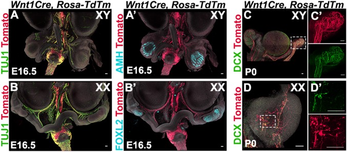
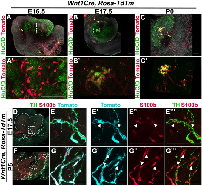
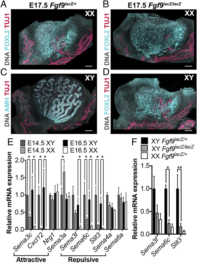
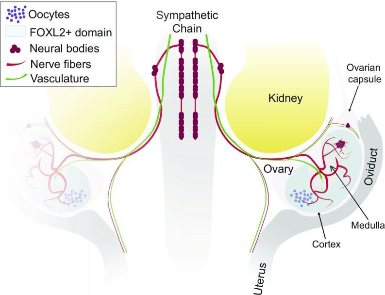
Testes and ovaries undergo sex-specific morphogenetic changes and adopt strikingly different morphologies, despite the fact that both arise from a common precursor, the bipotential gonad. Previous studies showed that recruitment of vasculature is critical for testis patterning. However, vasculature is not recruited into the early ovary. Peripheral innervation is involved in patterning development of many organs but has been given little attention in gonad development. In this study, we show that while innervation in the male reproductive complex is restricted to the epididymis and vas deferens and never invades the interior of the testis, neural crest-derived innervation invades the interior of the ovary around E16.5. Individual neural crest cells colonize the ovary, differentiate into neurons and glia, and form a dense neural network within the ovarian medulla. Using a sex-reversing mutant mouse line, we show that innervation is specific to ovary development, is not dependent on the genetic sex of gonadal or neural crest cells, and may be blocked by repressive guidance signals elevated in the male pathway. This study reveals another aspect of sexually dimorphic gonad development, establishes a precise timeline and structure of ovarian innervation, and raises many questions for future research.

Keywords: innervation; neural crest; organogenesis; ovary; testis.


Conflict of interest statement

The authors declare no conflict of interest.

Figures

Fig. 1.
Fig. 1.
Innervation reaches the gonad/mesonephros complex at E15.5. Whole-mount fluorescent immunostaining. (A and B) Urogenital complexes from E15.5 XX (A) or XY (B) embryos. The bladder was removed at dissection for better visualization. To best visualize the gonads, the XX sample (A) was imaged from the dorsal side, and the XY sample (B) was imaged from the ventral side. (CF) E15.5 gonads from XX (C and D) or XY (E and F) embryos imaged from the dorsal (C and E) or ventral (D and F) side. (C′) Magnified views (1 and 2) of the areas outlined in C. E′ and F′ are magnified views of the areas outlined in E and F. Samples were stained for the pan-neuronal marker TUJ1 (red, AF) and neural body marker HuC/D (green, AF). XX samples were stained for the granulosa cell marker FOXL2 to label the ovary (cyan, C and D), and XY samples were stained for the Sertoli cell marker AMH to label the testis (cyan, E and F). All samples were counterstained with Hoechst nuclear dye (grayscale). a, adrenal gland; k, kidney; m, mesonephros; o, ovary; p, pelvis; sc, sympathetic chain; t, testis. (Scale bars, 100 µm.)
Fig. 2.
Fig. 2.
Innervation invades the interior of the ovary but not the testis. (AD) E16.5 XY (A and C) or XX (B and D) gonads imaged from the ventral (A and B) or dorsal (C and D) side. (E and F) E18.5 XY (E) or XX (F) gonads imaged from the dorsal side. E′ and F′ are magnified views of the areas outlined in E and F that represent the Top and Bottom (28 µm deeper) optical sections from the Z-stacks used to generate maximum-intensity projections shown in E and F. (G and H) P0 XY (G) or XX (H) gonads imaged from the dorsal side. (IK) P5 XY (I) or XX (J and K) gonads imaged from the dorsal (I and J) or the ventral side (K). I′, J′, and K′ are magnified views of TUJ1 staining from the areas outlined in I, J, and K. Note that TUJ1 also stains Sertoli cells at some stages (I and I′). (LO) E17.5 (L and M) and P0 (N and O) XX gonads stained for the pan-neuronal marker TUJ1 (red, AO) and neural body marker HuC/D (green, LO). M and O are higher-magnification images of the areas outlined in L and N. Note that HuC/D also stains oocytes (LO and SI Appendix, Fig. S3). HuC/D staining in the oocyte is distinct from the stain observed in neural bodies: white arrowheads in O point to TUJ1-/HuC/D+ oocytes, and white arrows in O point to TUJ1+/HuC/D+ neural cell bodies. XY samples were stained for the Sertoli cell marker AMH (cyan, A, C, E, G, and I), and XX samples were stained for the granulosa cell marker FOXL2 (cyan, B, D, F, H, J, and K). All samples were counterstained with Hoechst nuclear dye (grayscale). cort, cortex; e, epididymis; ed, efferent ductules; h, hilus; m, mesonephros; med, medulla; o, ovary; t, testis; tt, testis tubules; vd, vas deferens. (Scale bars, 100 µm.)
Fig. 3.
Fig. 3.
Gonad innervation is neural crest-derived. (A and B) Lineage-trace of neural crest-derived neurons in whole urogenital complexes from XY (A) and XX (B) Wnt1Cre; Rosa-tdTomato embryos at E15.5. (C and D) Lineage-trace of neural crest-derived progenitors in gonads from XY (C) and XX (D) Wnt1Cre; Rosa-tdTomato neonates (P0). C′ and D′ are magnified views in single channels of the areas outlined in C and D. Samples were stained for red fluorescent protein (RFP) to detect tdTm expression (red, A and D), the pan-neuronal marker TUJ1 (green, A and B), and the pan-neuronal marker DCX (green, C and D). XY samples were stained for the Sertoli cell marker AMH to label the testis (cyan, A′), and XX samples were stained for the granulosa cell marker FOXL2 to label the ovary (cyan, B′). All samples were counterstained with Hoechst nuclear dye (grayscale). (Scale bars, 100 µm.)
Fig. 4.
Fig. 4.
Neural crest-derived progenitors colonize the ovary and differentiate into neurons and glia. (AC) Lineage-trace of neural crest-derived progenitors in gonads from XX Wnt1Cre; Rosa-tdTomato embryos at E16.5 (A), E17.5 (B), and P0 (C). A′, B′, and C′ are magnified views of the areas outlined in A, B, and C. Samples were stained for red fluorescent protein (RFP) to detect tdTm expression (red), the neural body marker HuC/D (green), and counterstained with Hoechst nuclear dye (grayscale). (DG) Lineage-trace of neural crest-derived neurons and glia in gonads from XX Wnt1Cre; Rosa-tdTomato embryos at E17.5 (D and E) and P5 (F and G). (E and G) Higher-magnification images of the areas outlined in D and F. Subsequent panels in each row show selected channels from the merged channels shown in E and G. Samples were stained for RFP to detect tdTm expression (cyan), the neural marker Tyrosine Hydroxylase (TH) (green), and the glial cell marker S100b (red). White arrowheads in E′ and G′ series point to tdTm+/S100b+/TH- cells in the ovary. (Scale bars, 100 µm.)
Fig. 5.
Fig. 5.
Neural crest migration is dependent on gonadal fate rather than genetic sex. (A and B) XX gonads from E17.5 Fgf9lacZ/+ (A) and Fgf9lacZ/lacZ (B) embryos. (C and D) XY gonads from E17.5 Fgf9lacZ/+ (C) or Fgf9lacZ/lacZ (D) embryos. All samples were stained for the pan-neuronal marker TUJ1 (red), and counterstained with Hoechst nuclear dye (grayscale). XX samples and Fgf9lacZ/lacZ XY samples were stained for the granulosa cell marker FOXL2 to label the ovary (cyan, A, B, and D). Fgf9lacZ/+ XY samples were stained with the Sertoli cell marker AMH to label the testis (cyan, C). (Scale bars, 100 µm.) (E) RT-qPCR analysis of relative mRNA expression of common neural crest attractive (Semaphorin 3c, Cxcl12, Neuregulin 1) or repulsive (Semaphorins 3a, 3f, 4a, 6a, 6c, and Slit3) cues in gonads from E14.5 (dashed bars) and E16.5 (solid bars) XY (black bars) or XX (white bars) embryos. Data were normalized to Gapdh expression. Values presented are the mean ± SEM of n = 4 pairs of gonads converted to fold changes compared with E14.5 XY samples for each gene. *P < 0.05 by two-tailed standard t test. (F) RT-qPCR analysis of relative mRNA expression of Semaphorins 3a, 6c, and Slit3 in gonads from E16.5 XY Fgf9lacZ/+ (black bars), XY Fgf9lacZ/lacZ (dashed bars), or XX Fgf9lacZ/+ (white bars) embryos. Data were normalized to Gapdh expression. Values presented are the mean ± SEM of n = 3 pairs of gonads converted to fold changes compared with XY Fgf9lacZ/+ samples for each gene. *P < 0.05; **P < 0.01 by two-tailed standard t test.
Fig. 6.
Fig. 6.
Innervation of the fetal mouse ovary. Neural crest-derived nerve fibers extend from the sympathetic chain posterior to the kidney and into the ovary. Neural crest-derived neural bodies are present in the anterior region of the ovarian capsule and also within the anterior region of the ovary. The ovarian neural network is dense in the ovarian medulla, a region without ovarian follicles, and extends projections across the FOXL2+ toward the ovarian cortex. While vasculature and innervation follow similar tracks to the ovary, they exhibit different patterns once inside the ovary.

Similar articles

Cited by

References

    1. Brennan J, Karl J, Capel B. Divergent vascular mechanisms downstream of Sry establish the arterial system in the XY gonad. Dev Biol. 2002;244:418–428. - PubMed
    1. Cool J, DeFalco TJ, Capel B. Vascular-mesenchymal cross-talk through Vegf and Pdgf drives organ patterning. Proc Natl Acad Sci USA. 2011;108:167–172. - PMC - PubMed
    1. Coveney D, Cool J, Oliver T, Capel B. Four-dimensional analysis of vascularization during primary development of an organ, the gonad. Proc Natl Acad Sci USA. 2008;105:7212–7217. - PMC - PubMed
    1. Combes AN, et al. Endothelial cell migration directs testis cord formation. Dev Biol. 2009;326:112–120. - PubMed
    1. Lei L, Spradling AC. Mouse oocytes differentiate through organelle enrichment from sister cyst germ cells. Science. 2016;352:95–99. - PMC - PubMed

Publication types

LinkOut - more resources