

Morphometric characteristics of the thoracοlumbar and lumbar vertebrae in the Greek population: a computed tomography-based study on 900 vertebrae-"Hellenic Spine Society (HSS) 2017 Award Winner"
- PMID: 30820461
- PMCID: PMC6381755
- DOI: 10.1186/s13013-019-0176-4
Morphometric characteristics of the thoracοlumbar and lumbar vertebrae in the Greek population: a computed tomography-based study on 900 vertebrae-"Hellenic Spine Society (HSS) 2017 Award Winner"
Abstract
Background: Vertebrae morphology appears to have genetic and ethnic variations. Knowledge of the vertebra and pedicle morphology is essential for proper selection and safe application of transpedicular screws. The aim of this study is to create a morphometric database for thoracolumbar and lumbar vertebrae (T9-L5) among individuals of both sexes in the Greek population.
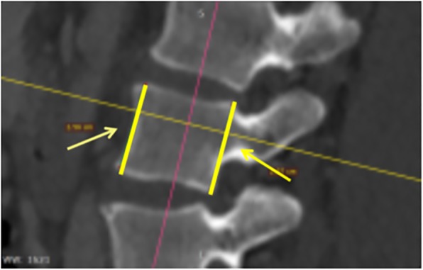
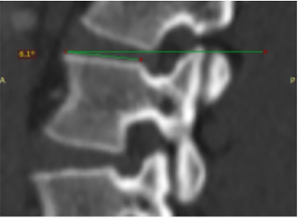
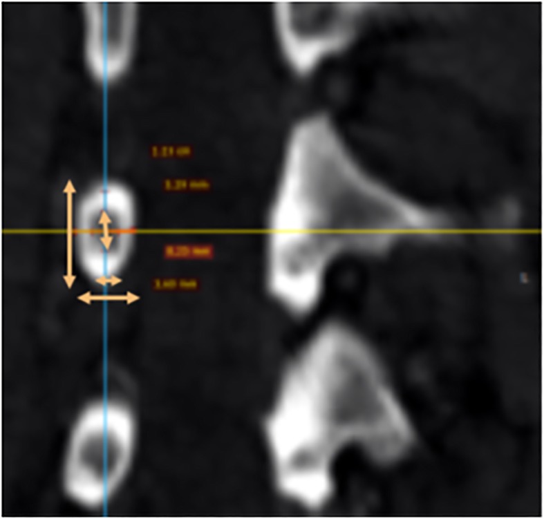
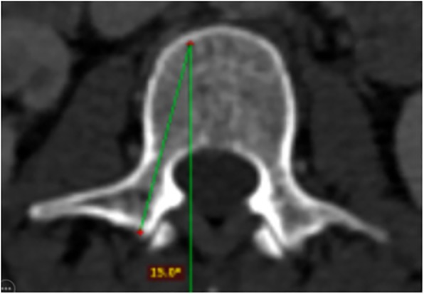
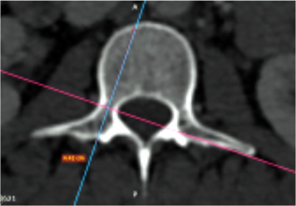
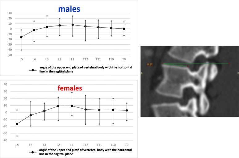
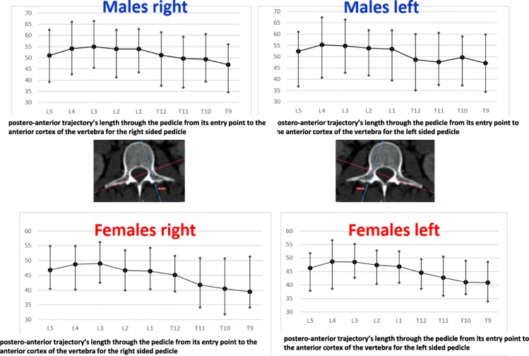
Material and methods: The morphometric dimensions of T9-L5 vertebrae on computed tomography (CT) scan images were measured in 100 adults (79 males and 21 females), without spinal pathology, age from 33 to 87 years old (mean 70 ± 8.73 years). The anterior vertebral body height (AVBH), the posterior vertebral body height (PVBH), the angle formed by the upper end plate of vertebral body and the horizontal line in the sagittal plane, the inner cancellous and outer cortical pedicle height and width, the angle formed by the longitudinal trajectory of the right- and left-sided pedicles and the midline anteroposterior axis of the vertebra (pedicle axis angle (PAA)), and the postero-anterior trajectory's length of the pedicle from the entry point to the anterior cortex of the vertebra (PTLP), for the right- and left-sided pedicles, were calculated. The Mann-Whitney U tests were conducted to compare the differences in various morphometric characteristics between sexes. The collected data were statistically analyzed using the SAS/STAT software 3.1.3 and SPSS version 22. The statistical significance was set at the level of p < 0.05. The intra- and inter-observer reliability of the measured parameters was also calculated.
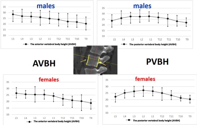
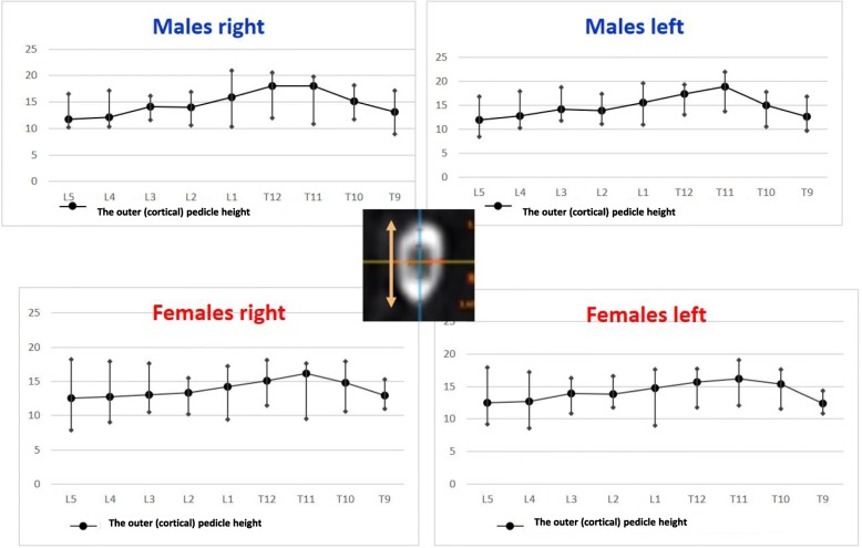
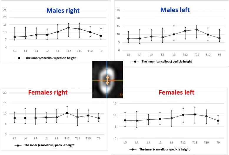
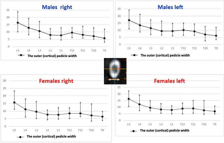
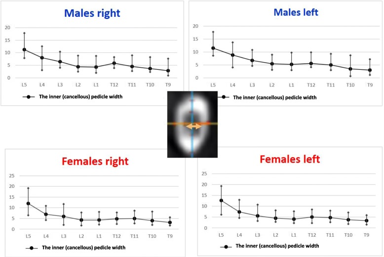
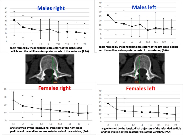
Results: The L5 vertebra had the maximum AVBH with a mean of 28.47 mm (SD ± 2.55 mm) in males and 26.48 mm (SD ± 1.61 mm) in females. The maximum PVBH in males was at L1 vertebra with a mean of 27.77 mm (SD ± 1.64 mm) and in females at L2 vertebral with a mean of 27.11 mm (SD ± 1.27 mm). Regarding the left pedicle dimensions, the maximum inner cancellous and outer cortical pedicle height was at T11 with a mean of 12.86 mm (SD ± 1.26 mm) and 18.82 mm (SD ± 1.37 mm) in males and 10.24 mm (SD ± 1.88 mm) and 16.19 mm (SD ± 3.27 mm) in females, respectively. The maximum inner cancellous and outer cortical pedicle width was at L5 with a mean of 11.57 mm (SD ± 1.97 mm) and 17.08 mm (SD ± 1.97 mm) in males and 10.24 mm (SD ± 1.88 mm) and 16.27 mm (SD ± 3.27 mm) in females, respectively. The largest PAA was found at the L5 with a mean angle of 26.23° (SD ± 2.65°) in males and 23.63° (SD ± 4.59°) in females, respectively. The maximum PTLP was found at the level of L4 with a mean of 55.31 mm (SD ± 4.52 mm) in males and 48.7 mm (SD ± 4.17 mm) in females, respectively. Regarding the right pedicle dimensions, the maximum inner cancellous and outer cortical pedicle height was found at T12 with a mean of 13.03 mm (SD ± 2.01 mm) and 18.01 mm (SD ± 1.56 mm) in males and 10.24 mm (SD ± 1.23 mm) and 16.14 mm (SD ± 1.23 mm) in females, respectively. The maximum inner cancellous and outer cortical pedicle width was at L5 with a mean of 11.3 mm (SD ± 2.86 mm) and 16.34 mm (SD ± 2.98 mm) in males and 12 mm (SD ± 3.18 mm) and 15.69 mm (SD ± 2.59 mm) in females, respectively. The greater PAA was at the L5 vertebral with a mean of 25.7° (SD ± 5.19°) in males and 25.56° (SD ± 5.31°) in females, respectively. The maximum PTLP was at the level of L3 with a mean of 54.86 mm (SD ± 3.18 mm) in males and 49.01 mm (SD ± 2.97 mm) in females, respectively. At all vertebrae, the only statistically significant difference (p < 0.0001) between the two sexes was the mean PTLP of the right and the left pedicle. The L5 vertebra was found to have the largest AVBH, PAA, and pedicle width in male and female populations.
Conclusions: This study provides a database of morphometric characteristics on thoracolumbar and lumbar vertebrae from T9 to L5 in the Greek population. This database may prove to be of great significance for forthcoming comparative studies. It can also serve as a basis in order to detect pathological changes in the spine and furthermore to plan operative interventions. It was found that the dimensions of thoracolumbar and lumbar vertebrae in the Greek population are sex-dependent. In the current study, vertebra and pedicle dimensions seem to have some similarities compared to other Western populations. However, in the thoracolumbar region, the pedicles of T9 and T10 may hardly accommodate a 4.00-mm pedicle screw given the narrow inner cancellous pedicle width. Importantly, the vertebra and pedicle dimensions measured in the current study can be used to guide the selection of transpedicular screws in the Greek population and to guide further research.
Keywords: A computed tomography-based study; Greek population; Lumbar vertebrae; Morphometric characteristics; Thoracolumbar vertebrae.
Conflict of interest statement
The study was approved by the Institutional Review Board of our Hospital. Excerpt of the minutes of the Tzaneio General Hospital IRB no: 40, 16/May/2017.not applicable.All authors declare that they have no competing interests.Springer Nature remains neutral with regard to jurisdictional claims in published maps and institutional affiliations.
Figures
Similar articles
-
A morphometric study of the thoracolumbar spine spinous process and lamina space in the Chinese.Folia Morphol (Warsz). 2021;80(3):665-674. doi: 10.5603/FM.a2020.0102. Epub 2020 Aug 26. Folia Morphol (Warsz). 2021. PMID: 32844385
-
A 3D-CT Study of the Cortical Bone Trajectory Screw Placement Parameters Based on Lumbar CT.Orthop Surg. 2024 Nov;16(11):2771-2780. doi: 10.1111/os.14202. Epub 2024 Aug 26. Orthop Surg. 2024. PMID: 39187426 Free PMC article.
-
Morphometric Analysis of the Lumbar Vertebrae Concerning the Optimal Screw Selection for Transpedicular Stabilization.Adv Exp Med Biol. 2019;1133:83-96. doi: 10.1007/5584_2018_324. Adv Exp Med Biol. 2019. PMID: 30680647
-
Morphometric Examination of the Larynx in Turkish Population.J Voice. 2024 Oct 23:S0892-1997(24)00343-6. doi: 10.1016/j.jvoice.2024.09.048. Online ahead of print. J Voice. 2024. PMID: 39448276 Review.
-
The Prevalence and Morphometry of the Atlas Vertebra Retrotransverse Foramen.Acta Med Acad. 2022 Dec;51(3):189-198. doi: 10.5644/ama2006-124.388. Epub 2022 Dec 15. Acta Med Acad. 2022. PMID: 36799311 Free PMC article.
Cited by
-
Computed Tomography-Based Morphometric Analysis of the Thoracolumbar Junction in the Young Turkish Population.Med Sci Monit. 2025 Jun 4;31:e948632. doi: 10.12659/MSM.948632. Med Sci Monit. 2025. PMID: 40465533 Free PMC article.
-
3D U-Net Neural Network Architecture-Assisted LDCT to Acquire Vertebral Morphology Parameters: A Vertebral Morphology Comprehensive Analysis in a Chinese Population.Calcif Tissue Int. 2024 Oct;115(4):362-372. doi: 10.1007/s00223-024-01255-8. Epub 2024 Jul 17. Calcif Tissue Int. 2024. PMID: 39017691
-
Morphological Parameters of the Thoracic Pedicle in an Asian Population: A Magnetic Resonance Imaging-Based Study of 3324 Pedicles.Global Spine J. 2021 May;11(4):437-441. doi: 10.1177/2192568220906137. Epub 2020 Feb 24. Global Spine J. 2021. PMID: 32875873 Free PMC article.
-
Needle path planning in semiautonomous and teleoperated robot-assisted epidural anaesthesia procedure: A proof of concept.Int J Med Robot. 2022 Dec;18(6):e2434. doi: 10.1002/rcs.2434. Epub 2022 Jun 29. Int J Med Robot. 2022. PMID: 35699156 Free PMC article.
-
Analysis of Screw/Pedicle-Width Ratio and Accuracy in Navigated Versus 3D-Controlled Fluoroscopy-Guided Pedicle Screw Placement.Global Spine J. 2025 May 19:21925682251343523. doi: 10.1177/21925682251343523. Online ahead of print. Global Spine J. 2025. PMID: 40387784 Free PMC article.
References
-
- Youkilis AS, Quint DJ, McGillicuddy JE, Papadopoulos SM. Stereotactic navigation for placement of pedicle screws in the thoracic spine. Neurosurgery. 2001;48(4):771–778. - PubMed
LinkOut - more resources
Full Text Sources
Research Materials