. 2019 Feb 25:2:76.
doi: 10.1038/s42003-019-0325-6. eCollection 2019.

Vascular RAGE transports oxytocin into the brain to elicit its maternal bonding behaviour in mice

Affiliations

Vascular RAGE transports oxytocin into the brain to elicit its maternal bonding behaviour in mice

Yasuhiko Yamamoto et al. Commun Biol. .

Abstract

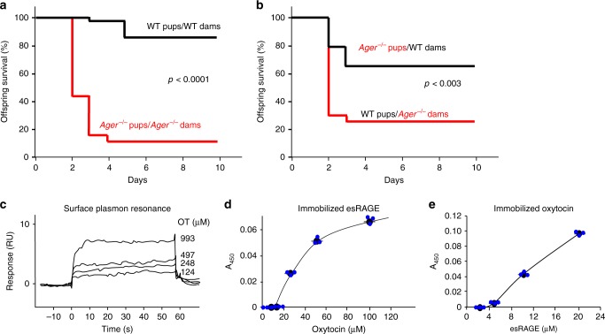
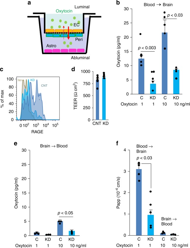
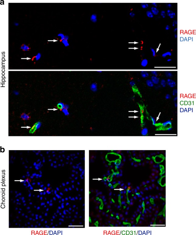
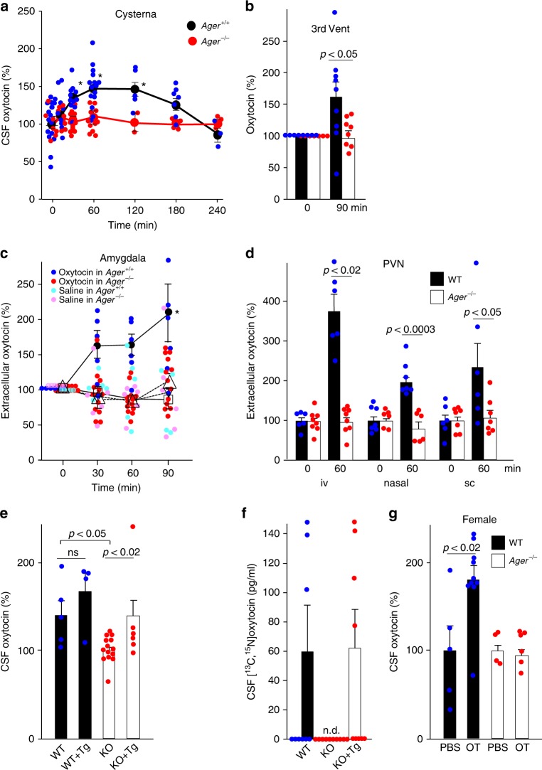
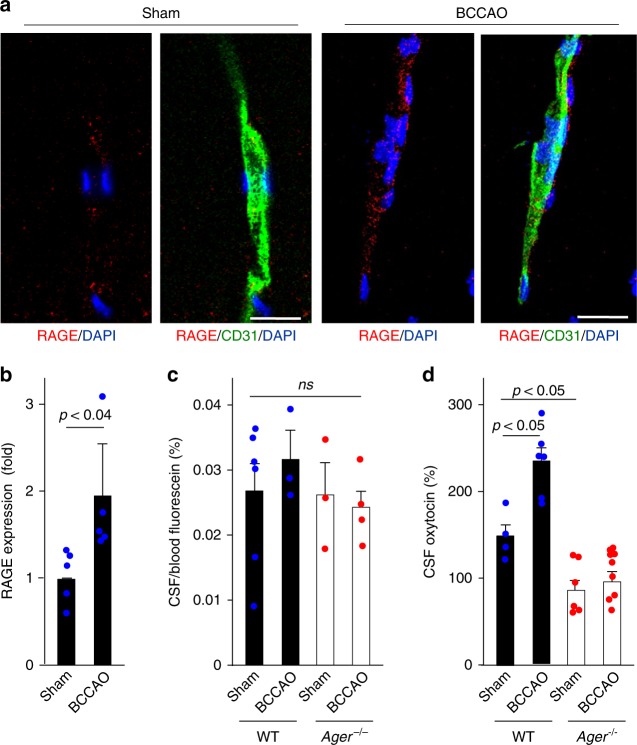
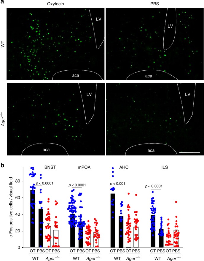
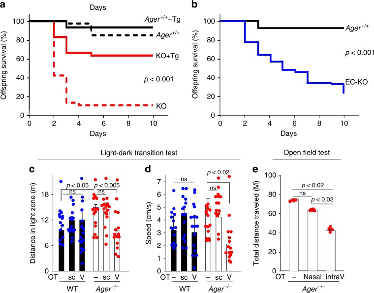
Oxytocin sets the stage for childbirth by initiating uterine contractions, lactation and maternal bonding behaviours. Mice lacking secreted oxcytocin (Oxt -/-, Cd38 -/-) or its receptor (Oxtr -/-) fail to nurture. Normal maternal behaviour is restored by peripheral oxcytocin replacement in Oxt -/- and Cd38 -/-, but not Oxtr -/- mice, implying that circulating oxcytocin crosses the blood-brain barrier. Exogenous oxcytocin also has behavioural effects in humans. However, circulating polypeptides are typically excluded from the brain. We show that oxcytocin is transported into the brain by receptor for advanced glycation end-products (RAGE) on brain capillary endothelial cells. The increases in oxcytocin in the brain which follow exogenous administration are lost in Ager -/- male mice lacking RAGE, and behaviours characteristic to abnormalities in oxcytocin signalling are recapitulated in Ager -/- mice, including deficits in maternal bonding and hyperactivity. Our findings show that RAGE-mediated transport is critical to the behavioural actions of oxcytocin associated with parenting and social bonding.


Conflict of interest statement

The authors declare no competing interests.

Figures

Fig. 1
Fig. 1
Offspring survival and RAGE-oxytocin interactions. Kaplan–Meier survival curves for offspring of WT (Ager+/+) and RAGE-null Ager−/− (knockout of the mouse gene for the receptor for AGEs) dams housed with their biological mothers (a; n = 40 WT pups/WT dams; 38 Ager−/−pups/Ager−/− dams), or transferred to different cages and housed with postpartum dams of the opposite genotype (b; n = 66 WT pups/Ager−/− dams; 66 Ager−/− pups/WT dams). P-values derived from log-rank calculations. c Surface plasmon resonance (BiaCore) assessment of oxytocin binding to immobilised human esRAGE. d, e Microtitre plate wells contain varying amounts of immobilised oxytocin (d) or esRAGE (e); added d esRAGE (1 μg/ml) or e oxytocin (100 μM) were detected immunochemically (n = 3). Values are mean ± SEM
Fig. 2
Fig. 2
RAGE transports oxytocin across an in vitro blood brain barrier (BBB). a A schematic diagram of the monkey BBB kit (PharmaCo-Cell). EC, monkey brain capillary endothelial cells; Peri, rat brain pericytes; Astro, rat astrocytes. The upper and the lower chambers represent the luminal (blood) and abluminal (brain extracellular space) sides, respectively. b Oxytocin (1 or 10 ng/ml) was added to the upper (luminal, blood) chambers of the model BBB system, and 3 h later oxytocin was quantified in the lower (abluminal, brain) chambers (n = 4–5). c Flow cytometry. The endothelial cells used in the upper chamber in a were treated with RAGE shRNA (knockdown, KD) or control (C or CNT) vectors to assess the effects of RAGE knockdown. Isotype control (Iso) instead of anti-RAGE antibody assesses background signal. d Transendothelial electrical resistance (TEER, Ωcm2) measures >150 Ωcm2 indicate integrity of the model BBB (n = 6). e Conversely, oxytocin was added to the abluminal chambers and its transport to the luminal chamber was quantified (n = 3). f The apparent permeability constants (Papp) for transfer were calculated from the distribution ratios across the chambers (n = 3–5). Values are mean ± SEM
Fig. 3
Fig. 3
RAGE in the brain vasculature. a, b Confocal microscopy. Sections of the hippocampus (a; CA1, stratum radiatum) and choroid plexus in the third ventricle (b) of WT male mice were immunostained with anti-RAGE and anti-CD31 antibodies; nuclei were stained with DAPI (Bar = 100 μm). Co-staining of RAGE (red, arrows) with CD31 (green) indicates that RAGE is present in some vascular endothelial cells
Fig. 4
Fig. 4
Transport of oxytocin into the brain. a Oxytocin concentrations in CSF from the cisterna magna after subcutaneous injection of 30 ng oxytocin in WT (Ager+/+) or Ager−/− male mice (n = 3–15/data point, *p < 0.05). b Oxytocin concentrations in the CSF of the third ventricles before and 90 min after subcutaneous administration of oxytocin (30 ng) (n = 7–9). c Oxytocin concentrations in microperfusates of the amygdala before and after intranasal (20 ng) oxytocin. Closed circles, oxytocin in WT mice; open triangles, oxytocin in Ager−/− mice; inverted open triangles, saline in WT mice; open squares, saline in Ager−/− mice (n = 4–13; *p < 0.05). d Oxytocin concentrations in microperfusates of the paraventricular nuclei (PVN) of WT mice before and 60 min after intravenous (iv), nasal (in) or subcutaneous (sc) administration of oxytocin (n = 6–8). e, f Transgenic (Tg) mice expressed human RAGE selectively in endothelial cells either on WT or Ager−/− (KO) backgrounds. Oxytocin concentrations in the CSF were measured 60 min after subcutaneous injections of oxytocin (e; n = 4–14) or [13C,15N]oxytocin (f; n = 9–10). g Oxytocin concentrations in the CSF of female mice were measured 60 min after subcutaneous injections of oxytocin (OT) (n = 4–10). n.d., not detected; ns, not significant. Values are mean ± SEM
Fig. 5
Fig. 5
Oxytocin transport after transient brain ischaemia. a Transient brain ischaemia was induced by 15 min of bilateral common carotid arteries occlusion (BCCAO) as described in Methods. RAGE and CD31 expression in vascular cells of the hippocampus were assessed 24 h after ischaemic insults; nuclei are stained with DAPI (Bar = 100 μm). b Quantitation of RAGE induction in CD31-positive endothelial cells (n = 5). c Fluorescein dye was used to check nonspecific vascular leakage and BBB damage following BCCAO (n = 3–6). ns, not significant. d Oxytocin concentrations in CSF from the cisterna magna of BCCAO or sham-operated WT (RAGE+/+) or Ager−/− male mice (n = 4–9). Values are mean ± SEM
Fig. 6
Fig. 6
Reporter assay for oxytocin activity in the brain. a 60 min after sc administration of oxytocin (OT) (30 ng) or PBS, c-Fos positive nuclei were visualised in the bed nucleus of the stria terminalis (BNST) of WT and Ager−/− male mice (LV, lateral ventricle; aca, anterior commissure, anterior part) (Bar = 100 μm). b Densities of c-Fos positive nuclei in the BNST, medial preoptic area (mPOA), centre of the anterior hypothalamic area (AHC), and intermediate lateral septal nucleus (ILS) regions of WT and Ager−/− mice (n = 12–56)
Fig. 7
Fig. 7
Offspring survival in Ager−/− mice and behavioural characteristic. a Survival curves for biological offspring of WT (Ager+/+) dams with (solid black line, n = 45) or without (black dashed line, n = 40) re-expressed human RAGE (Tg). Survival curves for biological offspring of Ager−/− (KO) dams with (red solid line, n = 71) or without (red dashed line, n = 38) re-expression of human RAGE. P < 0.001. b Survival curves for offspring of dams deficient for RAGE in endothelial cells (EC-KO, n = 76) and WT (Ager+/+) dams (solid black line, n = 45). P < 0.001. P-values derived from log-rank calculations. c, d During light-dark transition tests, the greater distances travelled (c) and greater average speed (d) in the light zone exhibited by Ager−/− male mice were both reduced by intraventricular (V) but not subcutaneous (sc) injection of oxytocin (OT) (n = 14–16). e In open field tests that assess anxiety in a new environment, the total distance travelled during the first 5 min was determined for Ager−/− male mice before and after intraventricular (intraV) (5 min, 0.1 ng/μl × 3 μl/min) or nasal (100 ng/ml × 20 μl) administration of oxytocin (OT) (n = 4–5). ns, not significant. Values are mean ± SEM

References

    1. Brunton PJ, Russell JA. The expectant brain: adapting for motherhood. Nat. Rev. Neurosci. 2008;9:11–25. doi: 10.1038/nrn2280. - DOI - PubMed
    1. Dulac C, O’Connell LA, Wu Z. Neural control of maternal and paternal behaviors. Science. 2014;345:765–770. doi: 10.1126/science.1253291. - DOI - PMC - PubMed
    1. Carter, C. S. Oxytocin and human evolution. Curr. Top. Behav. Neurosci. 35, 291–319 (2018). - PubMed
    1. Castel M, Gainer H, Dellmann HD. Neuronal secretory systems. Int. Rev. Cytol. 1984;88:303–459. doi: 10.1016/S0074-7696(08)62760-6. - DOI - PubMed
    1. Jin D, et al. CD38 is critical for social behaviour by regulating oxytocin secretion. Nature. 2007;446:41–45. doi: 10.1038/nature05526. - DOI - PubMed

Publication types

Substances