

Nephrotic syndrome in a dish: recent developments in modeling in vitro
- PMID: 30820702
- PMCID: PMC7316697
- DOI: 10.1007/s00467-019-4203-8
Nephrotic syndrome in a dish: recent developments in modeling in vitro
Abstract
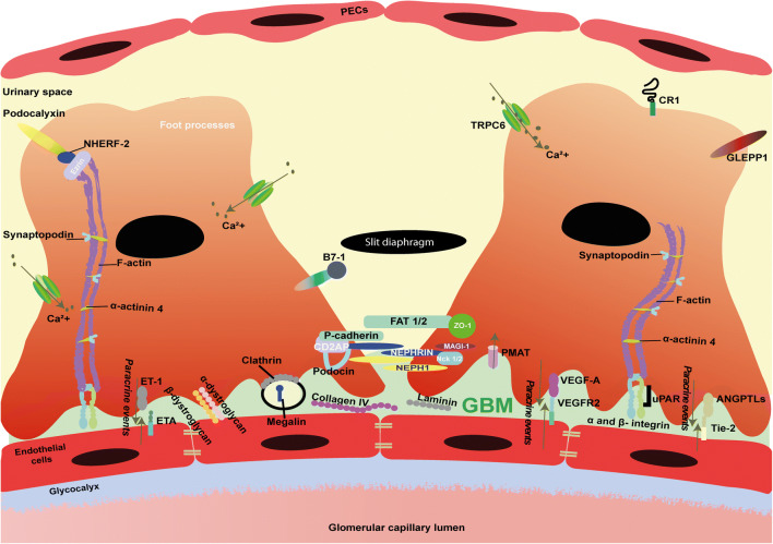
Nephrotic syndrome is a heterogeneous disease, and one of the most frequent glomerular disorders among children. Depending on the etiology, it may result in end-stage renal disease and the need for renal replacement therapy. A dysfunctional glomerular filtration barrier, comprising of endothelial cells, the glomerular basement membrane and podocytes, characterizes nephrotic syndrome. Podocytes are often the primary target cells in the pathogenesis, in which not only the podocyte function but also their crosstalk with other glomerular cell types can be disturbed due to a myriad of factors. The pathophysiology of nephrotic syndrome is highly complex and studying molecular mechanisms in vitro requires state-of-the-art cell-based models resembling the in vivo situation and preferably a fully functional glomerular filtration barrier. Current advances in stem cell biology and microfluidic platforms have heralded a new era of three-dimensional (3D) cultures that might have the potential to recapitulate the glomerular filtration barrier in vitro. Here, we highlight the molecular basis of nephrotic syndrome and discuss requirements to accurately study nephrotic syndrome in vitro, including an overview of specific podocyte markers, cutting-edge stem cell organoids, and the implementation of microfluidic platforms. The development of (patho) physiologically relevant glomerular models will accelerate the identification of molecular targets involved in nephrotic syndrome and may be the harbinger of a new era of therapeutic avenues.
Keywords: Glomerular filtration barrier; Kidney; Nephrotic syndrome; Organoids; Podocytes; Stem cells.
Conflict of interest statement
The authors declare that they have no conflict of interest.
Figures
References
-
- Noone DG, Iijima K, Parekh R. Idiopathic nephrotic syndrome in children. Lancet. 2018;392:61–74. - PubMed
-
- Kriz W, Hackenthal E, Nobiling R, Sakai T, Elger M, Hahnel B. A role for podocytes to counteract capillary wall distension. Kidney Int. 1994;45:369–376. - PubMed
-
- Zou J, Yaoita E, Watanabe Y, Yoshida Y, Nameta M, Li H, Qu Z, Yamamoto T. Upregulation of nestin, vimentin, and desmin in rat podocytes in response to injury. Virchows Arch. 2006;448:485–492. - PubMed
-
- Singh A, Satchell SC, Neal CR, McKenzie EA, Tooke JE, Mathieson PW. Glomerular endothelial glycocalyx constitutes a barrier to protein permeability. J Am Soc Nephrol. 2007;18:2885–2893. - PubMed
Publication types
MeSH terms
LinkOut - more resources
Full Text Sources
