. 2019 Jul:317:100-109.
doi: 10.1016/j.expneurol.2019.02.008. Epub 2019 Feb 26.

Dim light at night impairs recovery from global cerebral ischemia

Affiliations

Dim light at night impairs recovery from global cerebral ischemia

Laura K Fonken et al. Exp Neurol. 2019 Jul.

Abstract

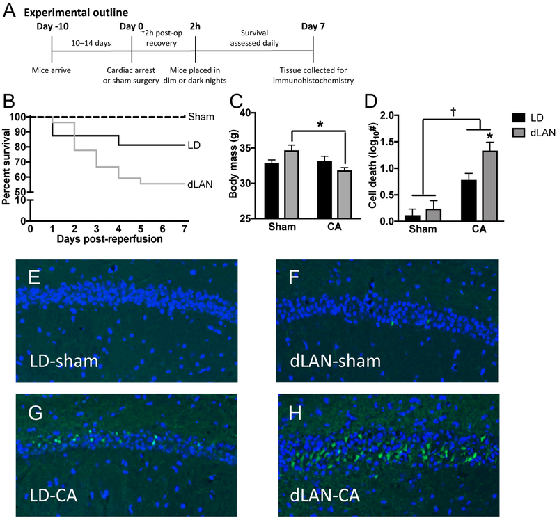
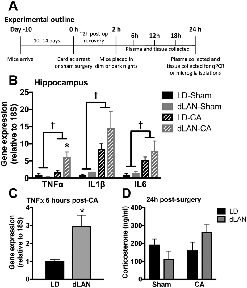
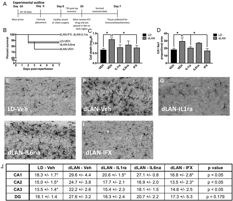
Nighttime lighting is one of the great conveniences of modernization; however, there is mounting evidence that inopportune light exposure can disrupt physiological and behavioral functions. Hospital patients may be particularly vulnerable to the consequences of light at night due to their compromised physiological state. Cardiac arrest/cardiopulmonary resuscitation (CA) was used to test the hypothesis in mice that exposure to dim light at night impairs central nervous system (CNS) recovery from a major pathological insult. Mice exposed to dim light at night (5 lx) had higher mortality in the week following cardiac arrest compared to mice housed in dark nights (0 lx). Neuronal damage was significantly greater in surviving mice exposed to dim light at night after CA versus those housed in dark nights. Dim light at night may have elevated neuronal damage by amplifying pro-inflammatory pathways in the CNS; Iba1 immunoreactivity (an indication of microglia activation) and pro-inflammatory cytokine expression were elevated in mice exposed to dim light at night post-CA. Furthermore, selective inhibition of IL-1β or TNFα ameliorated damage in mice exposed to dim light at night. The effects of light at night on CA outcomes were also prevented by using a wavelength of nighttime light that has minimal impact on the endogenous circadian clock, suggesting that replacing broad-spectrum nighttime light with specific circadian-inert wavelengths could be protective. Together, these data indicate that exposure to dim light at night after global cerebral ischemia increases neuroinflammation, in turn exacerbating neurological damage and potential for mortality.

Keywords: Cardiac arrest; Circadian; Cytokines; Light pollution; Microglia; Neuroinflammation.


Conflict of interest statement

CONFLICT OF INTEREST STATEMENT. The authors have no conflicts of interest to report. All authors concur with the submission of this manuscript and none of the data have been previously reported or are under consideration for publication elsewhere.

Figures

Figure 1.
Figure 1.. Dim light at night (dLAN) impairs recovery from a cardiac arrest and cardiopulmonary resuscitation (CA).
(a) Experimental outline for figure 1. Groups included n = 8 – 27 starting and n = 8 – 15 surviving; see supplemental Table 1). (b) Kaplan Meier curve illustrates short-term mortality during the first week following cardiac arrest/CPR (CA). (c) dLAN exposure reduced body mass in mice that underwent the CA procedure. (d) dLAN exacerbates CA induced neuronal damage in the hippocampus, as indicated by increased Fluoro-JadeC (FJC) staining relative to LD. Representative FJC stained sections (20X) from the CA1 region of the hippocampus: (e) Sham-dark night (LD-sham) (f) Sham-dim light at night (dLAN-sham) (g) CA-dark night (CA-LD) and (h) CA-dLAN at one week following the CA/sham procedure. Data were analyzed using a two-way ANOVA with Tukey’s post hoc test. The data in panel b represents mean±SEM; the dagger represents a significant main effect (p<0.05) between sham and CA, while the asterick indicates a significant difference (p<0.05) between CA-LD and CA-dLAN.
Figure 2.
Figure 2.. CA-induced pro-inflammatory cytokine elevations are exacerbated in the hippocampus of dLAN mice.
(a) Outline for experiments in figure 2 (n = 6 – 8/group; see supplemental table 1). (b) Hippocampal TNFα, IL1β, and IL6 mRNA concentrations are increased 24 h following CA relative to sham; furthermore, TNFα expression is potentiated by exposure to dLAN as compared to LD following CA in mice. (c) TNFα expression is elevated as early as 6 hours post-CA in the CA-dLAN group versus the CA-LD group (n = 4/group), this time point coincides with ~4 hours of exposure to dLAN. (d) Serum corticosterone concentrations at 24h were not altered by either CA or light at night (n = 6 – 8/group). There were no significant group differences in IL1β or IL6 at this early time point. Gene expression was assessed via RT-PCR and is presented as fold increase relative to a housekeeping gene (18S). The data are graphed as mean±SEM; a dagger symbol represents a significant main effect (p<0.05) between sham and CA, while an asterisk indicates a significant difference (p<0.05) between CA-LD and CA-dLAN.
Figure 3.
Figure 3.. Selective inhibition of specific pro-inflammatory cytokines attenuates inflammation and neuronal cell death following CA and dLAN.
(a) Experimental outline for figure 3 (n = 8 – 10/group, see supplemental table 1). (b) Kaplan Meier curve of mice treated with TNFα, IL1β or IL6 inhibitors 2 h following CA and directly prior to placement in dLAN. (c) Treatment of mice in the dLAN group with the TNFα or IL1β inhibitor reduced neuronal damage (FJC stain) and (d) inhibited proportional area of Iba1 staining at one week after CA relative to the dLAN vehicle treated mice. Representative photomicrographs of microglial staining with Iba1 in the CA1 region of the hippocampus (20X) in mice treated with (e) LD-Veh (f) dLAN-Veh (g) dLAN-IL1ra (h) dLAN-IL6ab or (i) dLAN-IFX. (j) Table provides proportional area of Iba1 staining in subregions of the hippocampus as an indication of microglial activation. The data in panels b, c, and i are provided as mean±SEM; Groups that differ from dLAN-VEH are marked with asterisks in the graphs (p<0.05).
Figure 4.
Figure 4.. Manipulation of lighting wavelength minimizes light at night induced damage following CA.
The experimental outline in Figure 4 followed the same timing as Figure 1a (n = 12/group, 6 – 10 surviving, see supplemental table 1). (a) Kaplan Meier curve illustrating survival of mice exposed to dark nights (LD), dim light at night (dLAN), or red light at night (rLAN) following CA. (b) the dLAN group had significantly greater neuronal damage (FJC staining) and (c) increased proportional area of Iba1 staining in the hippocampus at one week after CA than either the CA-LD or CA-rLAN groups; the CA-LD or CA-rLAN groups did not differ significantly in either of these measures. Representative photomicrographs of FJC staining in the CA1 region of the hippocampus (20X) one week after CA in the (d) LD, (e) dLAN, and (f) rLAN groups. Representative photomicrographs of Iba1 staining in the CA1 region of the hippocampus (20X) one week after CA in the (g) LD, (h) dLAN, and (i) rLAN groups. (j) Table providing proportional area of Iba1 staining in subregions of the hippocampus as an indication of microglial activation. (k) Gene expression of TNFα, IL1β, and IL6 is significantly elevated in the hippocampus 24 h following CA in the CA-dLAN group relative to the rLAN group and CA-LD groups (n = 7 – 8/group). Gene expression was assessed via qPCR and is presented as fold increase relative to a housekeeping gene (18S). The data are provided as mean±SEM; an asterisk represents a significant difference between dLAN and both other groups (p<0.05), whereas a pound sign (#) indicates the dLAN group only significantly differs from the rLAN group (p<0.05).

References

    1. Arruda AP, Milanski M, Coope A, Torsoni AS, Ropelle E, Carvalho DP, Carvalheira JB, Velloso LA, 2011. Low-grade hypothalamic inflammation leads to defective thermogenesis, insulin resistance, and impaired insulin secretion. Endocrinology 152, 1314–1326. - PubMed
    1. Ayalon L, Borodkin K, Dishon L, Kanety H, Dagan Y, 2007. Circadian rhythm sleep disorders following mild traumatic brain injury. Neurology 68, 1136–1140. - PubMed
    1. Bedrosian TA, Fonken LK, Nelson RJ, 2016. Endocrine Effects of Circadian Disruption. Annu Rev Physiol 78, 109–131. - PubMed
    1. Bedrosian TA, Galan A, Vaughn CA, Weil ZM, Nelson RJ, 2013a. Light at night alters daily patterns of cortisol and clock proteins in female Siberian hamsters. Journal of neuroendocrinology 25, 590–596. - PubMed
    1. Bedrosian TA, Weil ZM, Nelson RJ, 2013b. Chronic dim light at night provokes reversible depression-like phenotype: possible role for TNF. Mol Psychiatry 18, 930–936. - PubMed

Publication types

MeSH terms