. 2019:22:101694.
doi: 10.1016/j.nicl.2019.101694. Epub 2019 Jan 29.

Glutamate weighted imaging contrast in gliomas with 7 Tesla magnetic resonance imaging

Affiliations

Glutamate weighted imaging contrast in gliomas with 7 Tesla magnetic resonance imaging

Andrew Neal et al. Neuroimage Clin. 2019.

Abstract

Introduction: Diffuse gliomas are incurable malignancies, which undergo inevitable progression and are associated with seizure in 50-90% of cases. Glutamate has the potential to be an important glioma biomarker of survival and local epileptogenicity if it can be accurately quantified noninvasively.

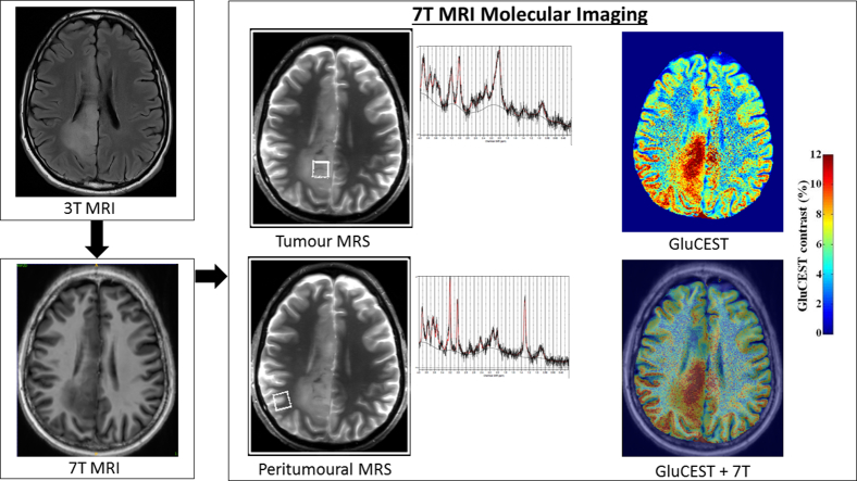
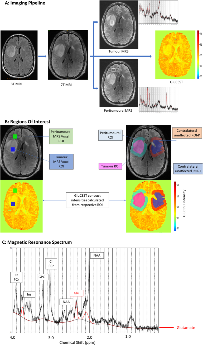
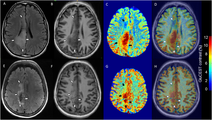
Methods: We applied the glutamate-weighted imaging method GluCEST (glutamate chemical exchange saturation transfer) and single voxel MRS (magnetic resonance spectroscopy) at 7 Telsa (7 T) to patients with gliomas. GluCEST contrast and MRS metabolite concentrations were quantified within the tumour region and peritumoural rim. Clinical variables of tumour aggressiveness (prior adjuvant therapy and previous radiological progression) and epilepsy (any prior seizures, seizure in last month and drug refractory epilepsy) were correlated with respective glutamate concentrations. Images were separated into post-hoc determined patterns and clinical variables were compared across patterns.

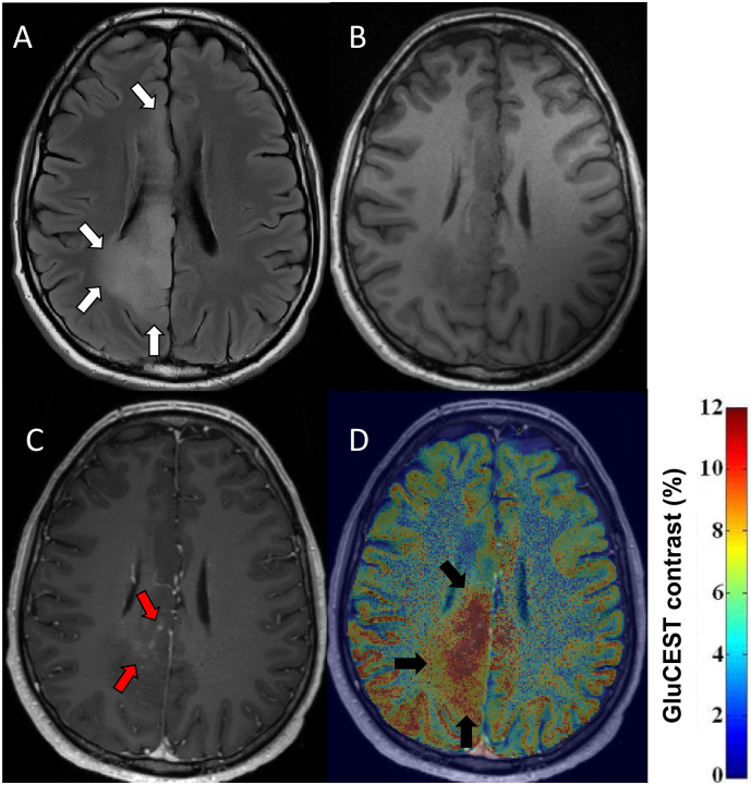
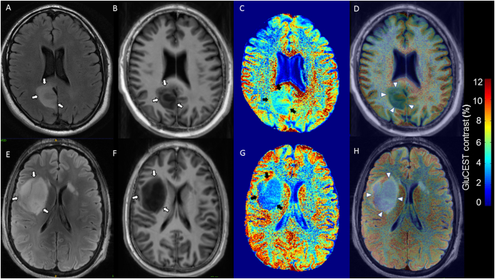
Results: Ten adult patients with a histo-molecular (n = 9) or radiological (n = 1) diagnosis of grade II-III diffuse glioma were recruited, 40.3 +/- 12.3 years. Increased tumour GluCEST contrast was associated with prior adjuvant therapy (p = .001), and increased peritumoural GluCEST contrast was associated with both recent seizures (p = .038) and drug refractory epilepsy (p = .029). We distinguished two unique GluCEST contrast patterns with distinct clinical and radiological features. MRS glutamate correlated with GluCEST contrast within the peritumoural voxel (R = 0.89, p = .003) and a positive trend existed in the tumour voxel (R = 0.65, p = .113).

Conclusion: This study supports the role of glutamate in diffuse glioma biology. It further implicates elevated peritumoural glutamate in epileptogenesis and altered tumour glutamate homeostasis in glioma aggressiveness. Given the ability to non-invasively visualise and quantify glutamate, our findings raise the prospect of 7 T GluCEST selecting patients for individualised therapies directed at the glutamate pathway. Larger studies with prospective follow-up are required.

Keywords: 7 T MRI; Epilepsy; Glioma; GluCEST; Glutamate; Seizure.


Figures

Unlabelled Image
Graphical abstract
Fig. 1
Fig. 1
7 T imaging protocol. Flow diagram describing imaging pipeline, regions of interest and magnetic resonant spectrum output. (A) 3 T axial image with the largest diameter of the FLAIR hyperintense lesion identified before 7 T MRI. Corresponding 7 T axial slice (axial GluCEST acquisition slab) determined with real-time visual comparison of 3 T and 7 T MRI. 5 mm GluCEST imaging performed at the GluCEST acquisition slab location. MRS performed in tumour and peritumoural tissue with a 15mm × 15mm × 15 mm voxel within the 5 mm thick GluCEST slab. (B) Regions of interest (ROI) drawn corresponding to i) tumour MRS voxel and peritumoural MRS voxel ii) tumour, iii) 1 cm rim around tumour border (peritumoural), iv) normal appearing brain in the contralateral hemisphere approximating a mirror image of the tumour ROI (contralateral unaffected ROI-T) and v) 1 cm rim around contralateral tumour ROI (contralateral unaffected ROI-P). Note, GluCEST contrast colour scheme adjusted from standard in this figure to allow better visualisation of ROIs. (C) Example of a magnetic resonance spectrum with the individual glutamate spectrum, outlined in red, showing three glutamate distinct peaks. Cr = creatine; Glu = glutamate; GPC = glycerophosphorylcholine; Ins = myo-inositol; NAA = N-acetyl aspartate; PCr = phosphocreatine.
Fig. 2
Fig. 2
GluCEST Contrast and gadolinium enhancing tumour. FLAIR, T1 weighted imaging with and without contrast and co-registered GluCEST imaging are presented for patient 4. WHO grade II oligodendroglioma IDH-mutant, 1p/19q codeleted, subsequent radiological progression and adjuvant therapy (temozolamide, radiotherapy). (A) 3 T FLAIR images in the axial GluCEST acquisition plane, tumour identified by white arrows. (B) 3 T T1 weighted images without gadolinium contrast in the axial GluCEST acquisition plane. (C) 3 T T1 weighted images with gadolinium contrast in the axial GluCEST acquisition plane with evidence of nodular, wispy mesial contrast enhancement (red arrows). (D) 3 T T1 weighted images with gadolinium co-registered with GluCEST contrast maps. Increased GluCEST contrast in a region overlapping, but extending beyond the area of gadolinium enhancement (black arrows).
Fig. 3
Fig. 3
Diffuse gliomas with low GluCEST contrast. FLAIR and T1 weighted images coregistered with GluCEST sequences revealing low tumour GluCEST contrast. (A–D) 37 year-old, seizure-free man with WHO grade II diffuse astrocytoma IDH-mutant (patient 3). No adjuvant therapy prior to 7 T MRI. (E–H) 30 year-old gentleman with tumour associated epilepsy, WHO grade II Diffuse astrocytoma IDH-mutant (patient 8). No adjuvant therapy prior to 7 T MRI. (A/E) = 3 T axial FLAIR (tumour identified by white arrows); (B/F) = 7 T axial T1 weighted imaging (tumour identified by white arrows); (C/G) = GluCEST contrast map (tumour identified by black arrows); (D) = Co-registered T1 and GluCEST (tumour identified by arrow heads); (H) = Co-registered FLAIR and GluCEST (tumour identified by arrow heads).
Fig. 4
Fig. 4
Tumours with heterogenous increased GluCEST contrast. FLAIR and T1 weighted images coregistered with GluCEST sequences revealing heterogenous increased tumour GluCEST contrast. (A–D) 33 year-old male, oligodendroglioma IDH-mutant, 1p/19q co-deleted at diagnosis, radiological progression of tumour and treatment with chemoradiotherapy prior to 7 T MRI (patient 4). (E–H) 55 year-old woman, oligodendroglioma IDH-mutant, 1p/19q co-deleted at diagnosis, radiological progression and treatment with chemotherapy prior to 7 T MRI (patient 7). (A) = 3 T axial FLAIR (tumour identified by white arrows); (E) = 7 T axial FLAIR (tumour identified by white arrows; (B/F) = 7 T axial T1 weighted imaging (tumour identified by white arrows); (C/G) = GluCEST contrast map (tumour identified by black arrows); (D/H) = Co-registered T1 and GluCEST (tumour identified by arrow heads).

References

    1. Balchandani P., Naidich T.P. Ultra-high-field MR neuroimaging. AJNR Am. J. Neuroradiol. 2015;36:1204–1215. - PMC - PubMed
    1. Bisdas S., Chadzynski G.L., Braun C., Schittenhelm J., Skardelly M., Hagberg G.E., Ethofer T., Pohmann R., Shajan G., Engelmann J., Tabatabai G., Ziemann U., Ernemann U., Scheffler K. MR spectroscopy for in vivo assessment of the oncometabolite 2-hydroxyglutarate and its effects on cellular metabolism in human brain gliomas at 9.4T. J. Magn. Reson. Imaging. 2016;44:823–833. - PubMed
    1. Buckingham S.C., Campbell S.L., Haas B.R., Montana V., Robel S., Ogunrinu T., Sontheimer H. Glutamate release by primary brain tumors induces epileptic activity. Nat. Med. 2011;17:1269–1274. - PMC - PubMed
    1. Buckner J.C., Shaw E.G., Pugh S.L., Chakravarti A., Gilbert M.R., Barger G.R., Coons S., Ricci P., Bullard D., Brown P.D., Stelzer K., Brachman D., Suh J.H., Schultz C.J., Bahary J.P., Fisher B.J., Kim H., Murtha A.D., Bell E.H., Won M., Mehta M.P., Curran W.J., Jr. Radiation plus procarbazine, CCNU, and vincristine in low-grade glioma. N. Engl. J. Med. 2016;374:1344–1355. - PMC - PubMed
    1. Cai K., Haris M., Singh A., Kogan F., Greenberg J.H., Hariharan H., Detre J.A., Reddy R. Magnetic resonance imaging of glutamate. Nat. Med. 2012;18:302–306. - PMC - PubMed

Publication types