

Validations of apomorphine-induced BOLD activation correlations in hemiparkinsonian rhesus macaques
- PMID: 30822717
- PMCID: PMC6396014
- DOI: 10.1016/j.nicl.2019.101724
Validations of apomorphine-induced BOLD activation correlations in hemiparkinsonian rhesus macaques
Abstract
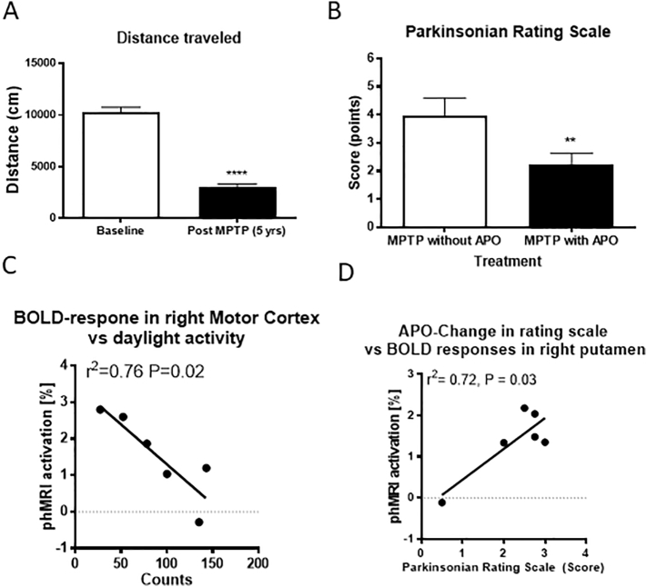
Identification of Parkinson's disease at the earliest possible stage of the disease may provide the best opportunity for the use of disease modifying treatments. However, diagnosing the disease during the pre-symptomatic period remains an unmet goal. To that end, we used pharmacological MRI (phMRI) to assess the function of the cortico-basal ganglia circuit in a non-human primate model of dopamine deficiency to determine the possible relationships between phMRI signals with behavioral, neurochemical, and histological measurements. Animals with unilateral treatments with the neurotoxin, 1-methyl-4-phenyl-1,2,3,6-tetrahydropyridine (MPTP), that expressed stable, long-term hemiparkinsonism were challenged with the dopaminergic receptor agonist, apomorphine, and structure-specific phMRI blood oxygen level-dependent (BOLD) activation responses were measured. Behavioral, histopathological, and neurochemical measurements were obtained and correlated with phMRI activation of structures of the cortico-basal ganglia system. Greater phMRI activations in the basal ganglia and cortex were associated with slower movement speed, decreased daytime activity, or more pronounced parkinsonian features. Animals showed decreased stimulus-evoked dopamine release in the putamen and substantia nigra pars compacta and lower basal glutamate levels in the motor cortex on the MPTP-lesioned hemisphere compared to the contralateral hemisphere. The altered neurochemistry was significantly correlated with phMRI signals in the motor cortex and putamen. Finally, greater phMRI activations in the caudate nucleus correlated with fewer tyrosine hydroxylase-positive (TH+) nigral cells and decreased TH+ fiber density in the putamen. These results reveal the correlation of phMRI signals with the severity of the motor deficits and pathophysiological changes in the cortico-basal ganglia circuit.
Keywords: Neurochemistry; Parkinson's disease; Pharmacological MRI; fMRI.
Copyright © 2019 The Authors. Published by Elsevier Inc. All rights reserved.
Figures


 ), the putamen shows areas of higher APO-induced BOLD activation along the rostral-caudal axis than in the unlesioned side (
 ). Sections were 3 mm in thickness through the putamen. *p < 0.05. (B) TH+ fiber density distribution along the anterior-posterior plane of the putamen in the MPTP-lesioned and the unlesioned hemispheres. (C) BOLD-responses in the right (MPTP-lesioned side) caudate were negatively correlated with the number of TH+ cells in the right substantia nigra. (D) BOLD-responses in the right motor cortex were negatively correlated with TH+ fibers in the right putamen.
References
-
- Alexander G.E., DeLong M.R., Strick P.L. Parallel organization of functionally segregated circuits linking basal ganglia and cortex. Annu. Rev. Neurosci. 1986;9:357–381. - PubMed
-
- Bifone A., Gozzi A. Neuromapping techniques in drug discovery: pharmacological MRI for the assessment of novel antipsychotics. Expert Opin. Drug Discovery. 2012;7:1071–1082. - PubMed
Publication types
MeSH terms
Substances
Grants and funding
LinkOut - more resources
Full Text Sources
Medical
Miscellaneous
