. 2019 Oct;86(4):460-470.
doi: 10.1038/s41390-019-0324-9. Epub 2019 Mar 1.

Increased miR-214 expression suppresses cell migration and proliferation in Hirschsprung disease by interacting with PLAGL2

Affiliations

Increased miR-214 expression suppresses cell migration and proliferation in Hirschsprung disease by interacting with PLAGL2

Liang Wu et al. Pediatr Res. 2019 Oct.

Abstract

Background: The miR-214 has been reported to be associated with various diseases, but its involvement in the pathophysiology of Hirschsprung disease (HSCR) is almost completely unexplored.

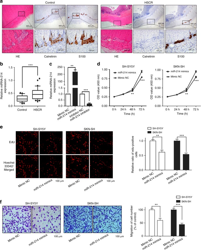
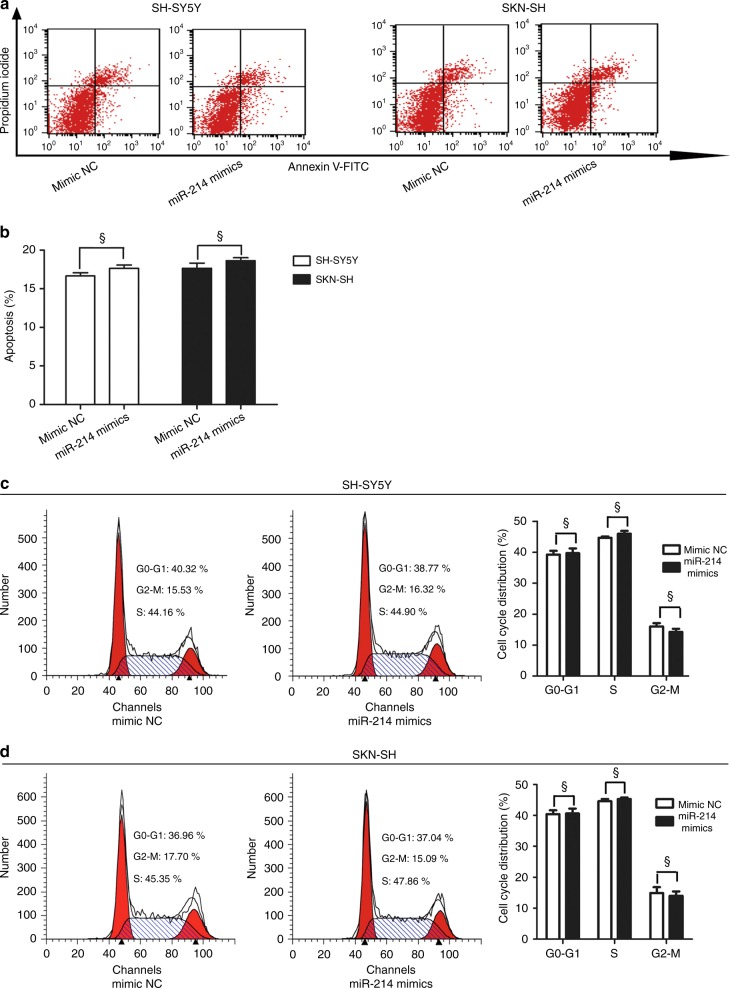
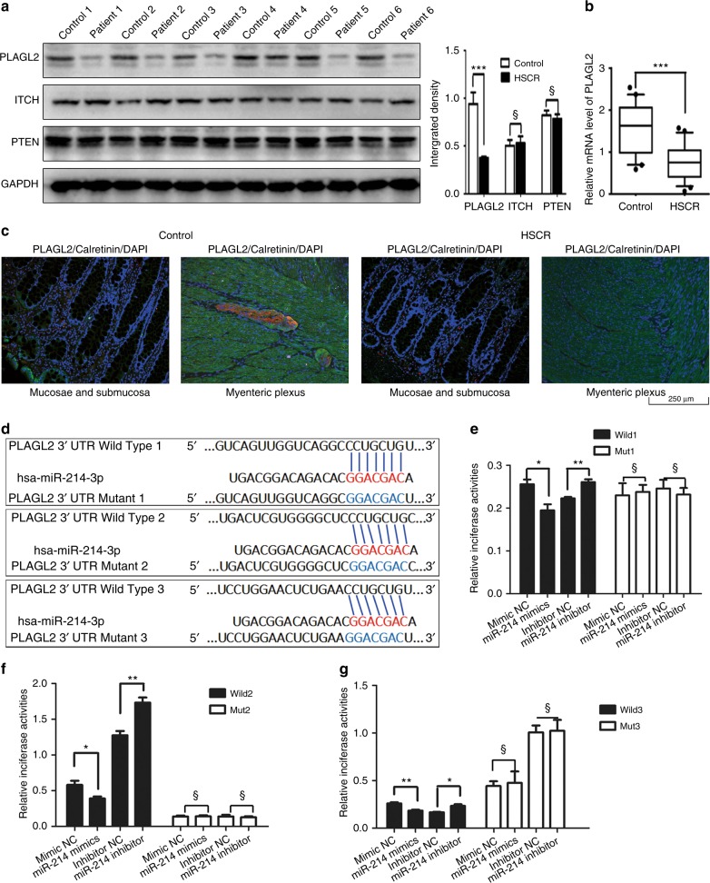
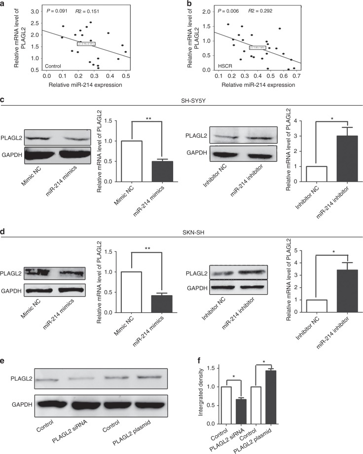
Methods: In our study, we conducted a series of experiments to unravel the biological role of miR-214 in the pathophysiology of HSCR. qRT-PCR and western blotting were utilized to investigate the relative expression levels of miR-214, mRNAs, and proteins of related genes in colon tissues from 20 controls without HSCR and 24 patients with HSCR. The potential biological role of miR-214 in two cell lines (SKN-SH and SH-SY5Y) was assessed using the CCK8 assay, EdU staining, transwell assay, and flow cytometry. The dual-luciferase reporter assay was used to confirm PLAGL2 as a common target gene of miR-214.

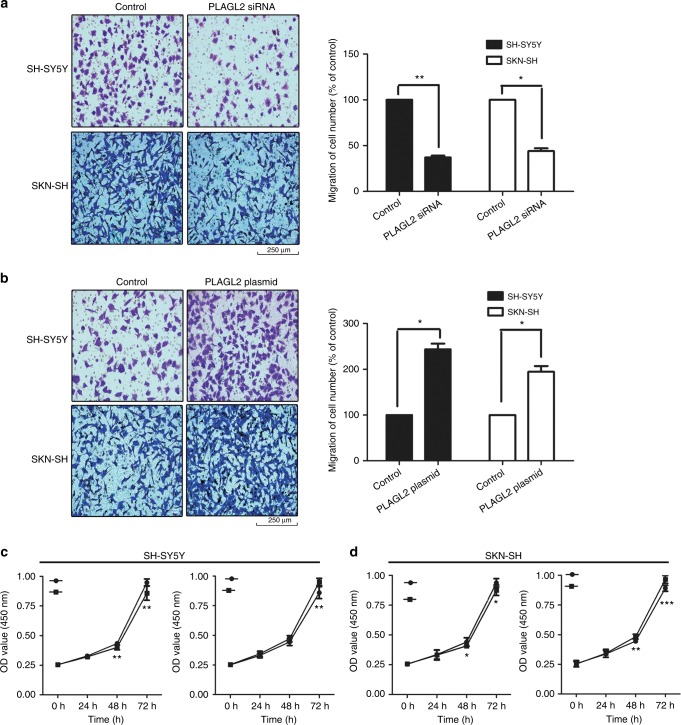
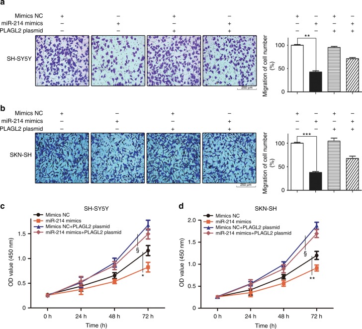
Results: All results suggested that miR-214 is upregulated in HSCR tissue samples compared with controls. Additionally, we found that miR-214 could inhibit cell proliferation and migration by directly downregulating the expression of PLAGL2, and the extent of the miR-214-mediated inhibitory effects could be rescued by a PLAGL2 overexpression plasmid.

Conclusion: Our results revealed that miR-214 is indeed involved in the pathophysiology of HSCR and suppresses cell proliferation and migration by directly downregulating PLAGL2 in cell models.


Conflict of interest statement

The authors declare no competing interests.

Figures

Fig. 1
Fig. 1
The overexpression of miR-214 in HSCR samples and the effects of the miR-214 mimics on cell growth and migration. * indicates a significant difference compared with control group (P < 0.05). ** indicates significant diferrences (P < 0.01) and *** indicates significant diferrences (P < 0.001). a the immunohistochemical staining for hematoxylin and eosin (HE), calretinin and S100 in HSCR and control group specimens. In control group specimens, calretinin staining for ganglion cells is positive and S100 staining highlighted negatively stained ganglion cells as a cell-shaped blank area. No calretinin staining was observed in HSCR tissues. Dense and prominent S100 immunostaining showed hypertrophic nerve trunks in HSCR specimens. b The expression of miR-214 in 24 HSCR cases was significantly higher compared to 20 matched controls (P = 0.0017). The expression level of miR-214 was analyzed using qRT-PCR and normalized with the data for U6. The data are presented as a box plot of the median and range of the log-transformed relative expression levels. c The relative expression level of miR-214 in the cells transfected with miR-214 mimics, miR-214 inhibitor, and the negative controls. df The effects of the miR-214 mimics on cell growth and migration. Representative images and the quantifications of migrated cells are shown. EdU proliferation assay analysis of the effect of miR-214 on the growth of SH-SY5Y and SKN-SH cells. The red fluorescent cells are in the S phase of mitosis, and the blue fluorescent cells represent all of the cells. The absorbance at 450 nm was presented as the mean ± SE. The migrated cells on the bottom of the membranes were visualized under 200× magnification and counted (six random views per well) with the ImageJ software
Fig. 2
Fig. 2
The effect of miR-214 mimics on apoptosis and cell cycle. § indicates no significant diferrence. ad A flow cytometry assay was performed to evaluate the effect of miR-214 mimics on apoptosis and cell cycle
Fig. 3
Fig. 3
PLAGL2 was a common target gene of miR-214. a, b The protein and RNA expression levels of PLAGL2, ITCH, and PTEN in human HSCR case and control tissues. c Immunofluorescence analysis of the PLAGL2 expression distribution. PLAGL2 (Alexa Fluor 488, green), Calretinin (Alexa Fluor 594, red). Nuclei were counterstained with DAPI. PLAGL2 was mainly expressed in lamina propria and the areas surrounding myenteric plexuses of control group specimens. The distribution of PLAGL2 in HSCR samples has no significant difference. d The sequence alignment of human miR-214 with the 3′ UTR of PLAGL2. There are three potential binding sites in the 3′ UTR region of PLAGL2 mRNA, The wild-type and mutated 3′ UTR sequences of PLAGL2 mRNA were called pGL3-PLAGL2-wild1–3 and pGL3-PLAGL2-mut1–3, respectively. eg The co-transfection of miR-214 mimics with pGL3-PLAGL2-wild1–3 remarkably suppressed the luciferase activity in the human SH-SY5Y cell line compared with the negative controls. However, no significant alteration between the luciferase activity of pGL3-PLAGL2-mut1–3 and negative controls was detected
Fig. 4
Fig. 4
The mRNA and protein expression levels of PLAGL2 were regulated by the miR-214. a, b The correlation was examined in HSCR cases, and it showed intense relationships between miR-214 and PLAGL2 (P = 0.006, R2 = 0.292), while a poor correlation was shown for the controls (P ≥ 0.05). c, d Two cell lines were transfected with 100 nM of miR-214 mimics, a miR-214 inhibitor and the respective controls for 48 h. Then, qRT-PCR and western blotting were performed to determine the mRNA and protein levels of PLAGL2. e, f The protein expression levels of PLAGL2 in the cells transfected with PLAGL2 siRNA, PLAGL2 plasmid, and the negative controls
Fig. 5
Fig. 5
The biological changes in cell migration and proliferation after treating two cell lines with PLAGL2 siRNA, PLAGL2 plasmid. ad The alterations of cell growth and migration after PLAGL2 siRNA and overexpression plasmid transfection in SH-SY5Y and SKN-SH cells
Fig. 6
Fig. 6
Co-transfection of the PLAGL2 plasmid could partially remove the miR-214 mimic-mediated suppression in cell proliferation and migration. ad The biological changes in migration and proliferation were partially reversed after co-transfection with miR-214 mimics and the PLAGL2 overexpression plasmid. We compared the co-transfected group with others and found that the migration and proliferation were partly reversed after co-transfection with miR-214 mimics and the PLAGL2 plasmid compared with the cells transfected with miR-214 mimics

References

    1. Pan Z, Li J. Advances in molecular genetics of Hirschsprung's disease. Anat. Rec. 2012;295:1628–1638. doi: 10.1002/ar.22538. - DOI - PubMed
    1. Emison Eileen Sproat, McCallion Andrew S., Kashuk Carl S., Bush Richard T., Grice Elizabeth, Lin Shin, Portnoy Matthew E., Cutler David J., Green Eric D., Chakravarti Aravinda. A common sex-dependent mutation in a RET enhancer underlies Hirschsprung disease risk. Nature. 2005;434(7035):857–863. doi: 10.1038/nature03467. - DOI - PubMed
    1. Carter TC, et al. Hirschsprung’s disease and variants in genes that regulate enteric neural crest cell proliferation, migration and differentiation. J. Hum. Genet. 2012;57:485–493. doi: 10.1038/jhg.2012.54. - DOI - PMC - PubMed
    1. McKeown SJ, et al. Hirschsprung disease: a developmental disorder of the enteric nervous system. Wiley Interdiscip. Rev. Dev. Biol. 2013;2:113–129. doi: 10.1002/wdev.57. - DOI - PubMed
    1. Tang W, et al. Suppressive action of miRNAs to ARP2/3 complex reduces cell migration and proliferation via RAC isoforms in Hirschsprung disease. J. Cell. Mol. Med. 2016;20:1266–1275. doi: 10.1111/jcmm.12799. - DOI - PMC - PubMed

Publication types