Review
. 2019 Feb 25;9(2):80.
doi: 10.3390/biom9020080.

Mechanisms of Spindle Positioning: Lessons from Worms and Mammalian Cells

Affiliations
Review

Mechanisms of Spindle Positioning: Lessons from Worms and Mammalian Cells

Sachin Kotak. Biomolecules. .

Abstract

Proper positioning of the mitotic spindle is fundamental for specifying the site for cleavage furrow, and thus regulates the appropriate sizes and accurate distribution of the cell fate determinants in the resulting daughter cells during development and in the stem cells. The past couple of years have witnessed tremendous work accomplished in the area of spindle positioning, and this has led to the emergence of a working model unravelling in-depth mechanistic insight of the underlying process orchestrating spindle positioning. It is evident now that the correct positioning of the mitotic spindle is not only guided by the chemical cues (protein⁻protein interactions) but also influenced by the physical nature of the cellular environment. In metazoans, the key players that regulate proper spindle positioning are the actin-rich cell cortex and associated proteins, the ternary complex (Gα/GPR-1/2/LIN-5 in Caenorhabditis elegans, Gαi/Pins/Mud in Drosophila and Gαi1-3/LGN/NuMA in humans), minus-end-directed motor protein dynein and the cortical machinery containing myosin. In this review, I will mainly discuss how the abovementioned components precisely and spatiotemporally regulate spindle positioning by sensing the physicochemical environment for execution of flawless mitosis.

Keywords: NuMA; actin cytoskeleton; dynein; microtubules; mitosis; myosin; spindle positioning.


Conflict of interest statement

The authors declare no conflict of interest.

Figures

Figure 1
Figure 1
Spindle positioning in Caenorhabditis elegans embryos and human cells. Images from the live-recording of the one-cell stage of C. elegans embryo expressing mCherry-tubulin (in grey) to label the microtubules are at various stages of cell cycle (A). Please note asymmetric spindle positioning in anaphase cells along the anterior-posterior axis, which would eventually lead to the unequal division of the one-cell embryo (not shown). Images from live-recording of HeLa cells stably expressing mCherry-H2B (in grey) to label the chromatin in interphase, and mitotic chromosomes at various stages of mitosis cultured on fibronectin-based L-shape micro-pattern. Please note that HeLa cell adapts the shape of the pattern during interphase; however, during metaphase when the cell is round spindle aligns along the longest axis. This causes chromosomes to separate along the longest axis when spindle elongates during anaphase (B).
Figure 2
Figure 2
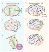
Cortically anchored ternary complex control spindle positioning in animal cells. Working model of spindle positioning in C. elegans embryo (A) and human cells (B). The ternary complex (LIN-5/GPR-1/2/Gα in C. elegans and NuMA/LGN/Gαi) is anchored below the cell cortex and recruits dynein motor protein complex. Dynein being a minus-end-directed microtubule-dependent motor complex attempts to move towards the centrosomes (shown by light brown arrow), but since it is anchored it instead pulls the astral microtubules resulting in the generation of the pulling forces towards the cell cortex (shown in black arrow). Please see text for more details.
Figure 3
Figure 3
Spindle positioning mechanisms in AB and EMS cells of C. elegans embryo. The mitotic spindle in AB and P1 cells at the two-cell stage of C. elegans embryo aligns parallel and perpendicular to the to the plane of AB-P1 cell contact respectively (A,B). Importantly, the alignment of the mitotic spindle and the cleavage plane in the AB cell is independent to GRP-1/2/dynein, and microtubules, but rather dependent on the unequal myosin-driven cortical flow that generates force to specify the position of the cleavage plane. Importantly, contact with an inert bead (shown in magenta) is sufficient to create the anisotropy in the myosin flow, and this creates an imbalance in the actomyosin-driven forces which defines the division site (C). Arrows represent the myosin flow, whereby it is slowest in the region where the AB cell contacts the bead (physical cue). Asymmetric division in EMS cell (shown on the right) into MS and E cell (DF). Prior to division, centrosome-nucleus complex rotates 90° under the influence of the Wnt signaling (shown in magenta) from the P2 cell (D,E). Wnt-signaling acts in concert with LIN-5/dynein pathway to instruct the mitotic spindle to align along the anterior-posterior axis. Please see text for more details.

References

    1. Walczak C.E., Heald R. Mechanisms of mitotic spindle assembly and function. Int. Rev. Cytol. 2008;265:111–158. - PubMed
    1. Jongsma M.L., Berlin I., Neefjes J. On the move: Organelle dynamics during mitosis. Trends Cell Biol. 2015;25:112–124. doi: 10.1016/j.tcb.2014.10.005. - DOI - PubMed
    1. Knoblich J.A. Mechanisms of asymmetric stem cell division. Cell. 2008;132:583–597. doi: 10.1016/j.cell.2008.02.007. - DOI - PubMed
    1. Gonczy P. Mechanisms of asymmetric cell division: Flies and worms pave the way. Nat. Rev. Mol. Cell Biol. 2008;9:355–366. doi: 10.1038/nrm2388. - DOI - PubMed
    1. Morin X., Bellaiche Y. Mitotic spindle orientation in asymmetric and symmetric cell divisions during animal development. Dev. Cell. 2011;21:102–119. doi: 10.1016/j.devcel.2011.06.012. - DOI - PubMed

Publication types