. 2019 Mar 1;11(3):533.
doi: 10.3390/nu11030533.

Hydrangea serrata (Thunb.) Ser. Extract Attenuate UVB-Induced Photoaging through MAPK/AP-1 Inactivation in Human Skin Fibroblasts and Hairless Mice

Affiliations

Hydrangea serrata (Thunb.) Ser. Extract Attenuate UVB-Induced Photoaging through MAPK/AP-1 Inactivation in Human Skin Fibroblasts and Hairless Mice

Hee-Soo Han et al. Nutrients. .

Abstract

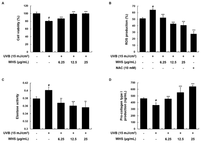
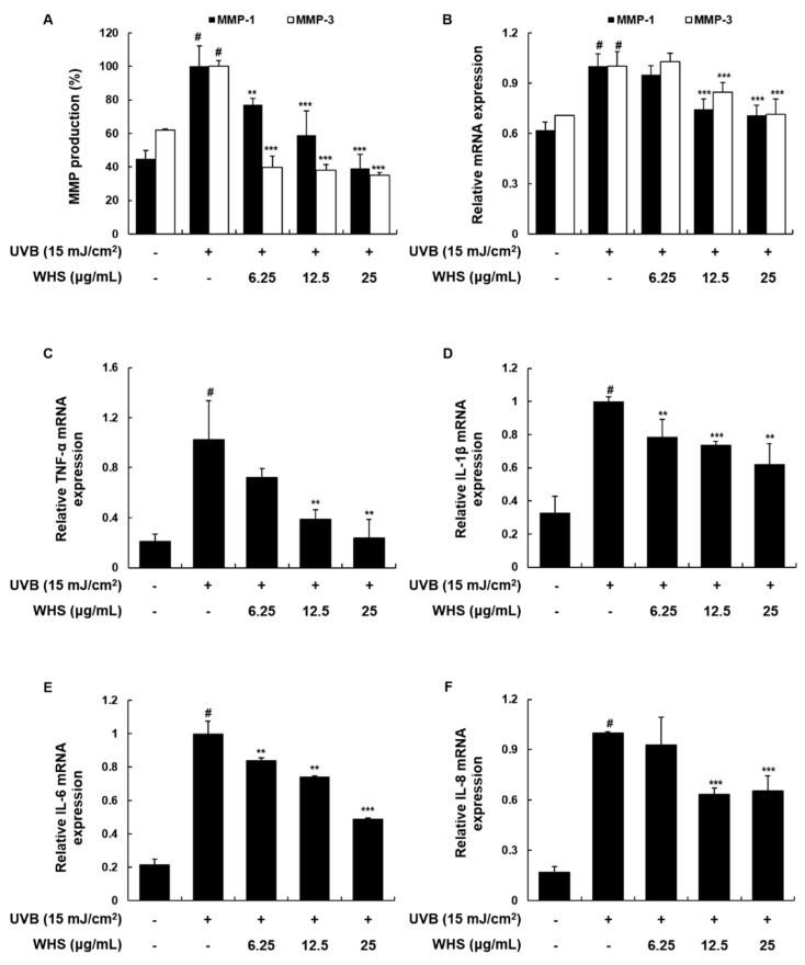
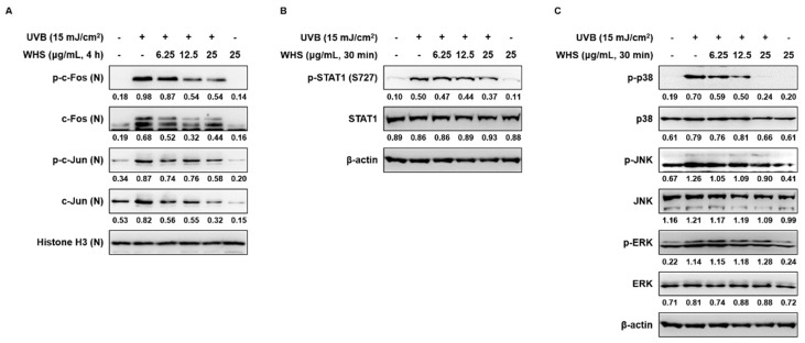
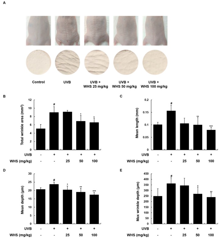
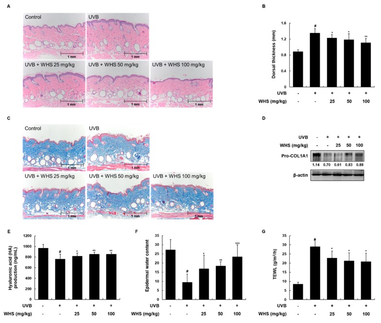
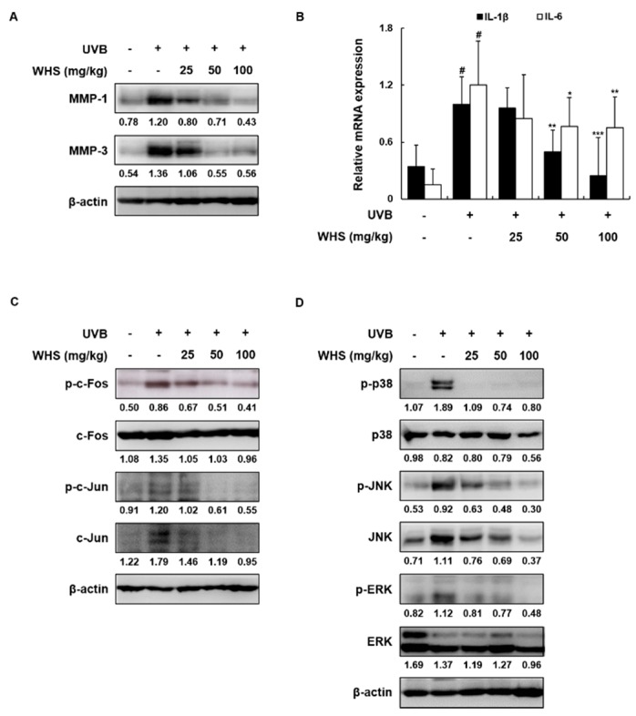
Skin photoaging is mainly caused by exposure to ultraviolet (UV) light, which increases expressions of matrix metalloproteinases (MMPs) and destroys collagen fibers, consequently inducing wrinkle formation. Nutritional factors have received scientific attention for use as agents for normal skin functions. The aim of this study was to investigate the effect of hot water extracts from the leaves of Hydrangea serrata (Thunb.) Ser. (WHS) against ultraviolet B (UVB)-induced skin photoaging and to elucidate the underlying molecular mechanisms in human foreskin fibroblasts (Hs68) and HR-1 hairless mice. WHS recovered UVB-reduced cell viability and ameliorated oxidative stress by inhibiting intracellular reactive oxygen species (ROS) generation in Hs68 cells. WHS rescued UVB-induced collagen degradation by suppressing MMP expression, and reduced the mRNA levels of inflammatory cytokines. These anti-photoaging activities of WHS were associated with inhibition of the activator protein 1 (AP-1), signal transduction and activation of transcription 1 (STAT1), and mitogen-activated protein kinase (MAPK) signaling pathways. Oral administration of WHS effectively alleviated dorsal skin from wrinkle formation, epidermal thickening, collagen degradation, and skin dehydration in HR-1 hairless mice exposed to UVB. Notably, WHS suppressed UVB activation of the AP-1 and MAPK signaling pathways in dorsal mouse skin tissues. Taken together, our data indicate that WHS prevents UVB-induced skin damage due to collagen degradation and MMP activation via inactivation of MAPK/AP-1 signaling pathway.

Keywords: AP-1; Hydrangea serrata (Thunb.) Ser.; MAPK; MMPs; collagen; photoaging; ultraviolet B.


Conflict of interest statement

The authors declare no conflict of interest.

Figures

Figure 1
Figure 1
Effects of Hydrangea serrata (Thunb.) Ser. (WHS) on cell proliferation, reactive oxygen species (ROS) generation, elastase activity, and procollagen type I production in UVB-induced Hs68 fibroblasts. Cells were exposed to UVB irradiation (15 mJ/cm2) and then treated with WHS (6.25, 12.5, or 25 μg/mL) for 24 h. (A) Cell viability was evaluated with an MTT assay. (B) After incubation, cells were stained with H2DCFDA (20 μM) for 30 min. ROS levels were evaluated by flow cytometry. N-acetyl-l-cystein (NAC) (10 mM), a common ROS inhibitor, was used as a positive control. (C) Cellular protein was prepared to examine elastase activity, determined using the elastase substrate STANA in WHS-treated cells. (D) The cell culture media were collected to determine the levels of procollagen type I. Values are expressed as means ± SD of three independent experiments. # p < 0.05 vs. the control group; ** p < 0.01, and *** p < 0.001 as compared to the UVB-irradiated group.
Figure 2
Figure 2
Effects of WHS on MMP-1/-3 protein and mRNA expression levels, and on mRNA expression of pro-inflammatory cytokines in UVB-exposed Hs68 fibroblasts. Cells were exposed to UVB irradiation (15 mJ/cm2) prior to WHS treatment (6.25, 12.5, or 25 μg/mL). (A) Following a 24 h incubation, the cell culture media were collected to determine MMP levels, using ELISA kits. (BF) Following a 3 h incubation, total cellular RNA was extracted from WHS-treated cells. The mRNA levels of MMPs, TNF-α, IL-1β, IL-6, and IL-8 were quantified by qRT-PCR and adjusted to GAPDH expression. Values are expressed as means ± SD of three independent experiments. # p < 0.05 vs. the control group; ** p < 0.01, and *** p < 0.001 as compared to the UVB-irradiated group.
Figure 3
Figure 3
Effects of WHS on the activator protein 1 (AP-1), signal transduction and activation of transcription 1 (STAT1), and mitogen-activated protein kinase (MAPK) signaling pathways in UVB-exposed Hs68 fibroblasts. Cells were irradiated with UVB (15 mJ/cm2) and then treated with WHS (6.25, 12.5, or 25 μg/mL) for 4 h (AP-1) or 30 min (STAT1 and MAPK). (A) Nuclear or (B,C) total cellular protein was prepared and resolved by SDS-PAGE, transferred to PVDF membranes, and detected with specific p-c-Fos (Ser32), p-c-Jun (Ser63), p-STAT1 (S727), p-p38, p-JNK, and p-ERK antibodies. Histone H3 and β-actin were used as internal controls for nuclear and whole-cell lysates, respectively. Presented data are the representative blots of three independent experiments.
Figure 4
Figure 4
Effects of WHS on skin wrinkle formation in UVB-irradiated hairless mice. HR-1 hairless mice were orally administrated WHS (25, 50, or 100 mg/kg) daily and exposed to UVB irradiation three times a week for 10 weeks. The skin replica samples of the dorsal areas were taken shortly after sacrifice. (A) Representative examples of external appearance of dorsal skin surface. The parameters for wrinkle formation, including (B) total wrinkle area, (C) mean length, (D) mean depth, and (E) maximum wrinkle depth, were obtained from the skin replica analysis. Values are expressed as means ± SD (n = 8). # p < 0.05 vs. the vehicle-treated control group; * p < 0.05, ** p < 0.01, and *** p < 0.001 as compared to the UVB only-treated group.
Figure 5
Figure 5
Effects of WHS on skin thickness and skin moisturizing factors in UVB-irradiated hairless mice. HR-1 hairless mice were orally administrated WHS (25, 50, or 100 mg/kg) daily and exposed to UVB irradiation three times a week for 10 weeks. (A) Skin tissues were stained with hematoxylin and eosin (H&E) to evaluate epidermal thickness and (B) the dorsal thickness of the skin of hairless mice was measured with a caliper before sacrifice. (C) Collagen fibers were identified with Masson’s trichrome staining. Skin tissues were homogenized for protein extraction. (D) Representative blots of pro-COL1A1 protein expression (n = 3). (E) Hyaluronic acid (HA) production was measured with the lysates using ELISA kits. (F) Skin hydration and (G) transepidermal water loss (TEWL) were measured in the back of the mice. Values are expressed as means ± SD (n = 8). # p < 0.05 vs the vehicle-treated control group; * p < 0.05, ** p < 0.01, and *** p < 0.001 as compared to the UVB only-treated group.
Figure 6
Figure 6
Effects of WHS on protein expression, pro-inflammatory cytokine mRNA levels, and the AP-1 and MAPK signaling pathways in UVB-irradiated hairless mice. HR-1 hairless mice were orally administrated WHS (25, 50, or 100 mg/kg) daily and exposed to UVB irradiation three times a week for 10 weeks. Skin tissues were homogenized for protein or RNA extraction. (A) Representative blots of MMP-1/-3 protein expression (n = 3). (B) The mRNA levels of IL-1β and IL-6 were quantified by qRT-PCR and adjusted to β-actin expression (n = 5). Representative blots of (C) AP-1 and (D) MAPK signaling pathway (n = 3). The protein lysates were resolved by SDS-PAGE, transferred to PVDF membranes, and detected with specific MMP-1/-3, p-c-Fos, p-c-Jun, p-p38, p-JNK, and p-ERK antibodies. β-actin was used as the internal control for total protein lysates. Values are expressed as means ± SD. # p < 0.05 vs. the vehicle-treated control group; * p < 0.05, ** p < 0.01, and *** p < 0.001 as compared to the UVB only-treated group.

Similar articles

Cited by

References

    1. Hwa C., Bauer E.A., Cohen D.E. Skin biology. Dermatol. Ther. 2011;24:464–470. doi: 10.1111/j.1529-8019.2012.01460.x. - DOI - PubMed
    1. Ganceviciene R., Liakou A.I., Theodoridis A., Makrantonaki E., Zouboulis C.C. Skin anti-aging strategies. Dermatoendocrinol. 2012;4:308–319. doi: 10.4161/derm.22804. - DOI - PMC - PubMed
    1. Subedi L., Lee T.H., Wahedi H.M., Baek S.H., Kim S.Y. Resveratrol-Enriched Rice Attenuates UVB-ROS-Induced Skin Aging via Downregulation of Inflammatory Cascades. Oxid Med. Cell. Longev. 2017;2017:8379539. doi: 10.1155/2017/8379539. - DOI - PMC - PubMed
    1. Bosch R., Philips N., Suarez-Perez J.A., Juarranz A., Devmurari A., Chalensouk-Khaosaat J., Gonzalez S. Mechanisms of Photoaging and Cutaneous Photocarcinogenesis, and Photoprotective Strategies with Phytochemicals. Antioxidants (Basel) 2015;4:248–268. doi: 10.3390/antiox4020248. - DOI - PMC - PubMed
    1. Kim H.K. Protective Effect of Garlic on Cellular Senescence in UVB-Exposed HaCaT Human Keratinocytes. Nutrients. 2016;8:464. doi: 10.3390/nu8080464. - DOI - PMC - PubMed