Review
. 2019 Mar 1;4(33):eaau6085.
doi: 10.1126/sciimmunol.aau6085.

Structure and function of the immune system in the spleen

Affiliations
Review

Structure and function of the immune system in the spleen

Steven M Lewis et al. Sci Immunol. .

Abstract

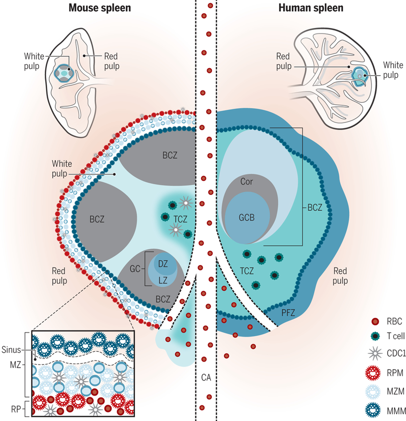
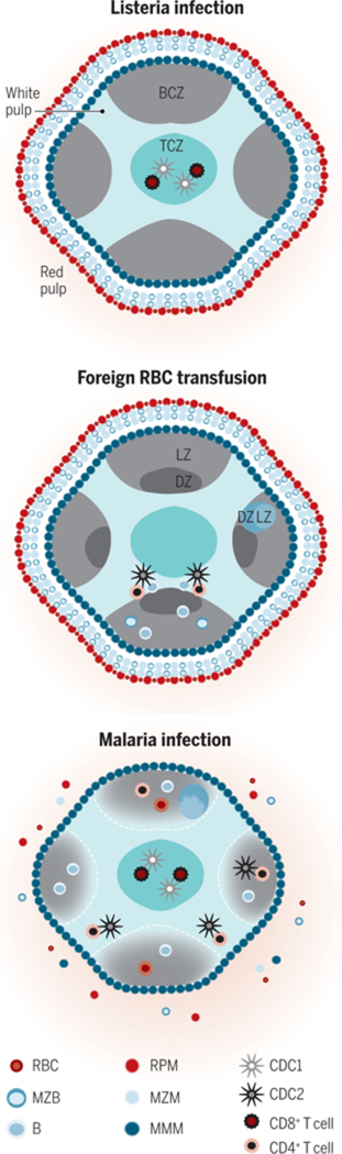
The spleen is the largest secondary lymphoid organ in the body and, as such, hosts a wide range of immunologic functions alongside its roles in hematopoiesis and red blood cell clearance. The physical organization of the spleen allows it to filter blood of pathogens and abnormal cells and facilitate low-probability interactions between antigen-presenting cells (APCs) and cognate lymphocytes. APCs specific to the spleen regulate the T and B cell response to these antigenic targets in the blood. This review will focus on cell types, cell organization, and immunologic functions specific to the spleen and how these affect initiation of adaptive immunity to systemic blood-borne antigens. Potential differences in structure and function between mouse and human spleen will also be discussed.


Figures

Figure 1
Figure 1. Mouse and human splenic immune cellular architecture at steady state.
There are structural differences between the murine (left) and human (right) splenic immune system, most notably, the organization of T cell zone (TCZ, turquoise; also known as PALS) and B cell zone (BCZ) follicles (gray and shades of blue, shown with light zone, LZ, and dark zone, DZ, organization in mouse spleen) within the WP and the border between the WP and RP, the MZ (marginal zone) in mouse or perifollicular zone (PFZ) in human (dark blue outer ring). Because applications of advanced imaging techniques to the human spleen have been limited, the extent to which the mouse MZ and human PFZ are analogous remains unknown. For example, the precise layering and composition of macrophage subsets in the MZ is known for mice (see bottom left box)—CD169+ MMMs (dark blue) form a concentric ring around the WP with MZMs (light blue) and MZB cells (darker blue)—but not for humans. In humans, MZB cells surround activated B cells, containing a GC (light blue in the human spleen on the right) and Corona (gray, “Cor”). The homeostatic location of dendritic cell (DC) subsets in mice is shown (with cDC2s in the bridging channel, BC, and cDC1s in the TCZ, MZ and RP, red pulp). Release of blood into the MZ of the WP from a central arteriole (CA) is shown.
Figure 2
Figure 2. The cellular organization of the murine WP is dynamic.
The cells of the TCZ (turquoise) are organized into CD8+ T cells (maroon) in the center and CD4+ T cells (orange) in the outer TCZ. This organization enables rapid formation of necessary cellular contacts, after migration within the WP, for the response to an immunologic insult. Shown are selected, dominant immune reactions in the spleen relevant to each insult. (A) Listeria induces rapid migration of cDC1s (silver) into the central TCZ where they present antigen and prime CD8+ T cells. (B) Transfusion of xenogeneic or allogeneic RBCs induces migration of cDC2s (black) to the outer TCZ, for CD4+ T cell priming and effective B cell antibody production (with the LZ, light zone, and DZ, dark zone, of a germinal center, GC, shown). (C) Malaria-infected RBCs induce a poorly coordinated and slow humoral immune response that includes disintegration of WP-RP border, MZ disruption and GC disorganization. After repeated infections, protective antibodies are ultimately produced.

References

    1. Bronte V, Pittet MJ, The spleen in local and systemic regulation of immunity. Immunity 39, 806–818 (2013). - PMC - PubMed
    1. van Krieken JH, te Velde J, Normal histology of the human spleen. Am J Surg Pathol 12, 777–785 (1988). - PubMed
    1. Nolte MA, Hoen EN, van Stijn A, Kraal G, Mebius RE, Isolation of the intact white pulp. Quantitative and qualitative analysis of the cellular composition of the splenic compartments. European journal of immunology 30, 626–634 (2000). - PubMed
    1. Khanna KM, Lefrancois L, Geography and plumbing control the T cell response to infection. Immunology and cell biology 86, 416–422 (2008). - PMC - PubMed
    1. Mebius RE, Kraal G, Structure and function of the spleen. Nature reviews. Immunology 5, 606–616 (2005). - PubMed

Publication types