. 2019 Apr 26;294(17):6772-6784.
doi: 10.1074/jbc.RA118.006642. Epub 2019 Mar 1.

A conserved and buried edge-to-face aromatic interaction in small ubiquitin-like modifier (SUMO) has a role in SUMO stability and function

Affiliations

A conserved and buried edge-to-face aromatic interaction in small ubiquitin-like modifier (SUMO) has a role in SUMO stability and function

Kiran Sankar Chatterjee et al. J Biol Chem. .

Abstract

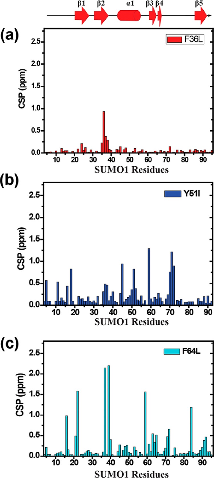
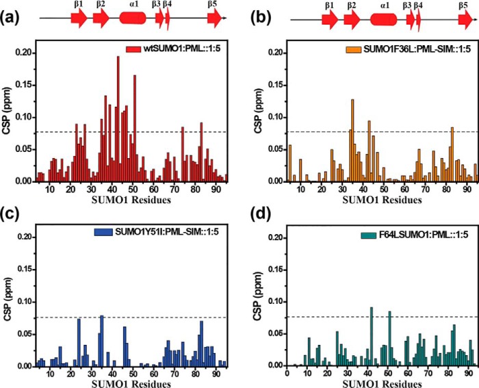
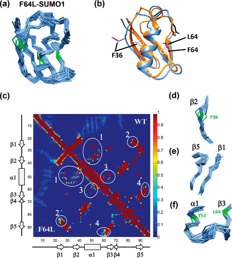
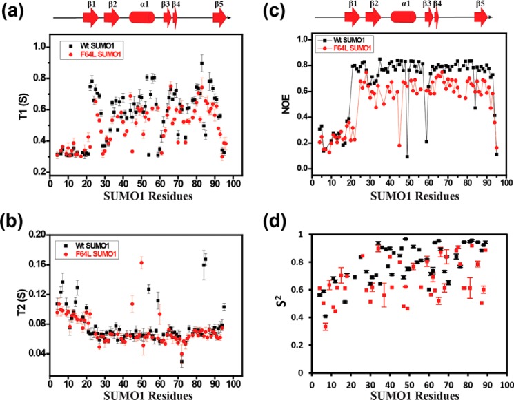
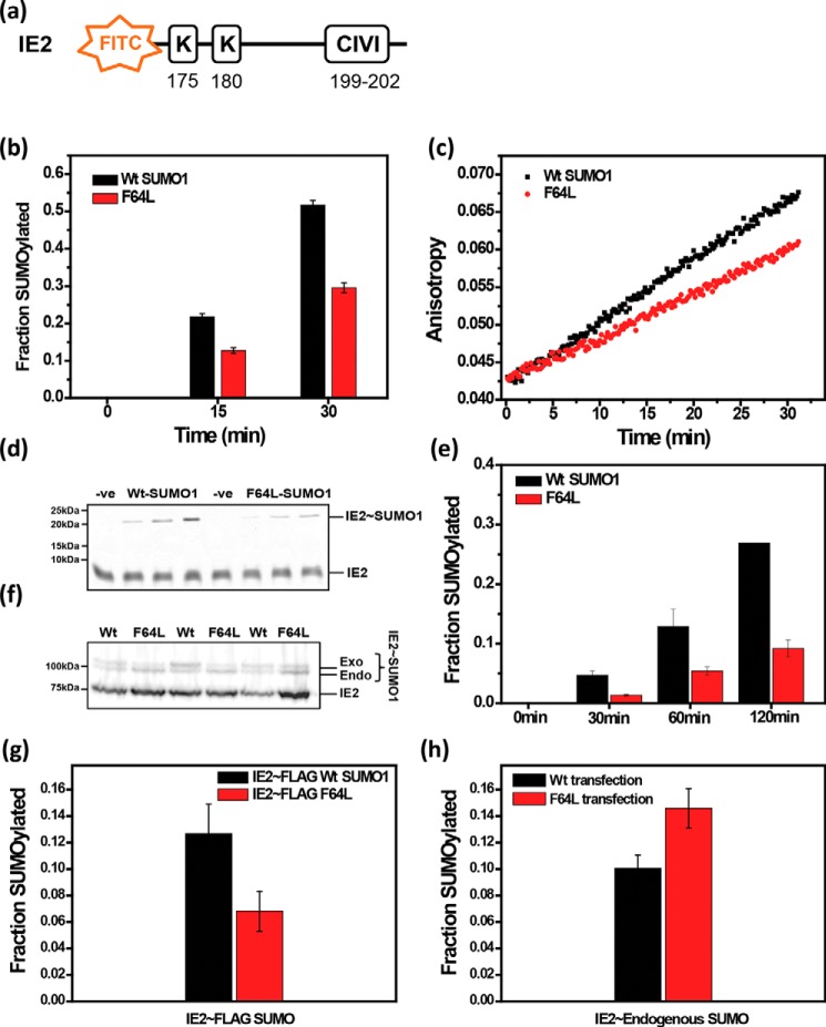
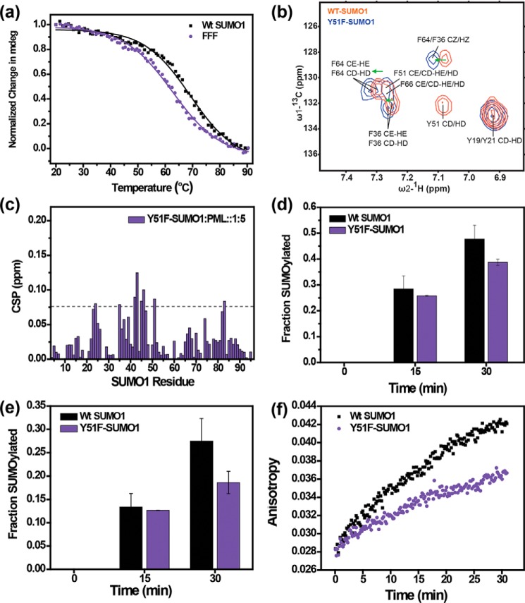
Aromatic amino acids buried at a protein's core are often involved in mutual paired interactions. Ab initio energy calculations have highlighted that the conformational orientations and the effects of substitutions are important for stable aromatic interactions among aromatic rings, but studies in the context of a protein's fold and function are elusive. Small ubiquitin-like modifier (SUMO) is a common post-translational modifier that affects diverse cellular processes. Here, we report that a highly conserved aromatic triad of three amino acids, Phe36-Tyr51-Phe64, is a unique SUMO signature that is absent in other ubiquitin-like homologous folds. We found that a specific edge-to-face conformation between the Tyr51-Phe64 pair of interacting aromatics is vital to the fold and stability of SUMO. Moreover, the noncovalent binding of SUMO-interacting motif (SIM) at the SUMO surface was critically dependent on the paired aromatic interactions buried at the core. NMR structural studies revealed that perturbation of the Tyr51-Phe64 conformation disrupts several long-range tertiary contacts in SUMO, leading to a heterogeneous and dynamic protein with attenuated SUMOylation both in vitro and in cells. A subtle perturbation of the edge-to-face conformation by a Tyr to Phe substitution significantly decreased stability, SUMO/SIM affinity, and the rate of SUMOylation. Our results highlight that absolute co-conservation of specific aromatic pairs inside the SUMO protein core has a role in its stability and function.

Keywords: SUMO-interacting motif (SIM); aromatic interactions; aromatic ring; aromatic triad; nuclear magnetic resonance (NMR); post-translational modification (PTM); protein dynamics; protein misfolding; protein structure; sumoylation.


Conflict of interest statement

The authors declare that they have no conflicts of interest with the contents of this article

Figures

Figure 1.
Figure 1.
a, multiple sequence alignment of SUMO homologs from human, mouse, and Arabidopsis. Conserved residues are highlighted in black, and the conserved aromatic triad is marked by red arrows. Solvent accessibility is shown at the bottom of alignment in a color scale from black to white, where black indicates solvent exposed and white represents buried residues. b, structure of SUMO1 (4WJO) (19) in blue and (c) structure of ubiquitin (1UBQ) (35) in green and are shown indicating the aromatic triad (FYF) in SUMO and the aliphatic side chains (red) (LIL) in the ubiquitin core at corresponding positions, respectively. d, a schematic representation of two predominant aromatic ring conformations, parallel offset stacking, and edge-to-face arrangement. e, zoomed in view at the SUMO hydrophobic core shows an edge-to-face interaction between Phe64 and Tyr51 aromatic moieties.
Figure 2.
Figure 2.
a, far UV circular dichroism spectra showing conserved secondary structure for wt and SUMO1 mutants. b, temperature melts for SUMO1 aromatic mutants. Change in mean ellipticity is normalized and plotted against temperature. c, urea-induced denaturation melt for each mutant. Normalized mean ellipticity shift is plotted against urea concentration. d, the plot of steady-state ANS fluorescence intensity upon binding with wt and mutant proteins.
Figure 3.
Figure 3.
CSP upon mutation plotted against individual residues of different SUMO mutants (a) F36L, (b) Y51I, (c) F64L. The chemical shift perturbations between the wt and mutants are calculated as CSP = [(δWtH − δmutantH)2 + ((δWtN − δmutantN)/5)2]1/2, where δH and δN are the chemical shift of the amide hydrogen and nitrogen, respectively.
Figure 4.
Figure 4.
CSP plotted against individual residues of different SUMO mutants. a, wt SUMO1; b, F36L; c, Y51I; d, F64L. The chemical shift perturbations between the free and PML-SIM bound forms are calculated as CSP = [(δ freeH − δboundH)2 + ((δfreeN − δboundN)/5)2]1/2, where δH and δN are the chemical shift of the amide hydrogen and nitrogen, respectively. The dashed line indicates mean ± S.D. of CSP values for wt, residues exhibiting CSPs above the dashed line are significantly perturbed and indicate the binding interface. The secondary structure alignment of SUMO1 against its sequence is provided on top.
Figure 5.
Figure 5.
Solution structure of F64L-SUMO1. a, the backbone Cα chain of the 20 lowest energy structures of F64L-SUMO1 is shown, colored in blue. Phe36-Tyr51-Phe64 backbone Cα atoms and bonds are colored green. b, the lowest energy structure of wt-SUMO1 (PDB 2N1V) and the mutant is compared. Ribbons are colored in orange and blue, for the wt-SUMO1 and F64L-SUMO1, respectively. The Phe36 and Phe64 side chains are shown in gray and purple for WT and mutant, respectively. c, the contact map of WT and F64L SUMO1 is shown in the same 2D plot, where WT is represented in the upper right, and the F64L is represented in the lower left region of the plot. The major differences between the contacts are circled and labeled. The areas 1, 2, and 4 in c are shown in d–f, respectively.
Figure 6.
Figure 6.
Comparison of 15N-H relaxation time constants T1 (a), T2 (b), heteronuclear NOE (c), and the order parameter (S2) (d) between WT (black) and mutant SUMO1 (red). Individual values are plotted against SUMO1 residues. The secondary structure alignment of SUMO1 against its sequence is provided on top of each plot.
Figure 7.
Figure 7.
SUMOylation assay with wt and F64L mutant SUMO1. a, schematic representation of the SUMOylation site and SIM in substrate IE2; b, in vitro SUMOylation level of IE2 quantified for wt SUMO1 and F64L mutant; c, fluorescence anisotropy of FITC-IE2 plotted against time from 0 to 30 min for wt and F64L. d, FITC imaged SDS-PAGE of SUMOylation between wt SUMO1 and F64L for SIM-less IE2 as a substrate. e, quantification of SUMOylation of SIM-less IE2 as a substrate. f, Western blotting image of in vivo IE2 SUMOylation. g, SUMOylation of IE2 in HEK 293T. The plot of the SUMOylated fraction of IE2 by exogenous wt and F64L-SUMO1 from three different replicates. (h) SUMOylation of IE2 by endogenous SUMO proteins plotted for wt-SUMO1 and F64L mutant.
Figure 8.
Figure 8.
Effect of tyrosine to phenylalanine substitution at Tyr51. a, temperature melt profile for wt and Y51F-SUMO1. Change in mean ellipticity is normalized and plotted against temperature. b, overlap of the assigned aromatic spectra of wt (red) and Y51F-SUMO1 (black). The green arrows show downfield peak shifts upon the Y51F mutation. c, the chemical shift perturbations of the Y51F-SUMO/PML-SIM interaction plotted against SUMO1 residues. d and e, in vitro SUMOylation level of wt-IE2 (d) and SIM-less IE2 (e) quantified for wt-SUMO1 and Y51F-SUMO1. f, fluorescence anisotropy of wt-FITC-IE2 plotted against time from 0 to 30 min for wt and Y51F-SUMO1.

References

    1. Hillier B. J., Rodriguez H. M., and Gregoret L. M. (1998) Coupling protein stability and protein function in Escherichia coli CspA. Fold. Des. 3, 87–93 10.1016/S1359-0278(98)00014-5 - DOI - PubMed
    1. Hong H., Park S., Jiménez R. H., Rinehart D., and Tamm L. K. (2007) Role of aromatic side chains in the folding and thermodynamic stability of integral membrane proteins. J. Am. Chem. Soc. 129, 8320–8327 10.1021/ja068849o - DOI - PubMed
    1. Frank B. S., Vardar D., and Buckley D. A., and McKnight C. J. (2002) The role of aromatic residues in the hydrophobic core of the villin headpiece subdomain. Protein Sci. 11, 680–687 10.1110/ps.22202 - DOI - PMC - PubMed
    1. Jiang X., Loo D. D., Hirayama B. A., and Wright E. M. (2012) The importance of being aromatic: π interactions in sodium symporters. Biochemistry 51, 9480–9487 10.1021/bi301329w - DOI - PMC - PubMed
    1. Bhattacharya S., and Ainavarapu S. R. K. (2018) Mechanical softening of a small ubiquitin-related modifier protein due to temperature induced flexibility at the core. J. Phys. Chem. B 122, 9128–9136 10.1021/acs.jpcb.8b06844 - DOI - PubMed

Publication types

LinkOut - more resources