. 2019 Apr 15;79(8):2031-2041.
doi: 10.1158/0008-5472.CAN-18-3259. Epub 2019 Mar 1.

MUC1-C Integrates Chromatin Remodeling and PARP1 Activity in the DNA Damage Response of Triple-Negative Breast Cancer Cells

Affiliations

MUC1-C Integrates Chromatin Remodeling and PARP1 Activity in the DNA Damage Response of Triple-Negative Breast Cancer Cells

Masaaki Yamamoto et al. Cancer Res. .

Abstract

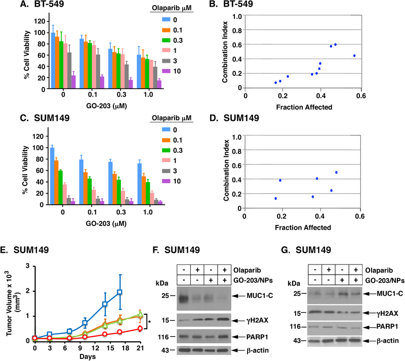
The oncogenic MUC1-C protein is overexpressed in triple-negative breast cancer (TNBC) cells and contributes to their epigenetic reprogramming and chemoresistance. Here we show that targeting MUC1-C genetically or pharmacologically with the GO-203 inhibitor, which blocks MUC1-C nuclear localization, induced DNA double-strand breaks and potentiated cisplatin (CDDP)-induced DNA damage and death. MUC1-C regulated nuclear localization of the polycomb group proteins BMI1 and EZH2, which formed complexes with PARP1 during the DNA damage response. Targeting MUC1-C downregulated BMI1-induced H2A ubiquitylation, EZH2-driven H3K27 trimethylation, and activation of PARP1. As a result, treatment with GO-203 synergistically sensitized both mutant and wild-type BRCA1 TNBC cells to the PARP inhibitor olaparib. These findings uncover a role for MUC1-C in the regulation of PARP1 and identify a therapeutic strategy for enhancing the effectiveness of PARP inhibitors against TNBC. SIGNIFICANCE: These findings demonstrate that targeting MUC1-C disrupts epigenetics of the PARP1 complex, inhibits PARP1 activity, and is synergistic with olaparib in TNBC cells.


Conflict of interest statement

Conflict of Interest: DK has equity interests in, serves as a member of the board of directors of and is a paid consultant to Genus Oncology. The other authors disclosed no potential conflicts of interest.

Figures

Figure 1.
Figure 1.. Nuclear localization of MUC1-C in the DDR.
A. Structure of the MUC1-C protein with the 58 aa extracellular domain (ED), 28 aa transmembrane domain (TM) and 72 aa cytoplasmic domain (CD). Highlighted is the aa sequence of the CD, which is an intrinsically protein (IDP). The CQC motif is necessary for MUC1-C homodimerization and nuclear localization, and is the target of the cell-penetrating peptide GO-203 (R9-CQCRRKN). B and C. BT-549 (B) and SUM149 (C) cells were left untreated or treated with 10 μM CDDP for 24 h. Total cell (left) and nuclear (right) lysates were analyzed by immunoblotting with antibodies against the indicated proteins. D and E. BT-549 (D) and SUM149 (E) cells were left untreated (Control) or treated with 10 FM CDDP for 24 h. The cells were stained with DAPI and analyzed for nuclear MUC1-C localization by confocal microscopy.
Figure 2.
Figure 2.. Silencing MUC1-C induces γH2AX.
A and B. BT-549 (A) and SUM149 (B) cells stably expressing a tet-MUC1shRNA were treated with 500 ng/ml DOX for 72 h. Total cell lysates were immunoblotted with antibodies against the indicated proteins. C and D. BT-549/tet-MUC1shRNA (C) and SUM149/tet-MUC1shRNA (D) cells were treated with 500 ng/ml DOX for 72 h and then 10 μM CDDP for 24 h. Lysates were subjected to immunoblot analysis. E and F. BT-549/tet-MUC1shRNA (E) and SUM149/tet-MUC1shRNA (F) cells were treated with 500 ng/ml DOX and/or 10 μM CDDP for 10 days. Colony formation was determined by crystal violet staining. The results are expressed as the mean±SD of three determinations. An asterisk denotes a p-value of <0.05.
Figure 3.
Figure 3.. Targeting MUC1-C with the GO-203 inhibitor attenuates nuclear localization of MUC1-C and induces γH2AX.
A-D. BT-549 (A,C) and SUM149 (B,D) cells were left untreated or treated with 1 μM GO-203 for 48 h and then without or with 10 μM CDDP for 24 h. Nuclear (A,B) and total (C,D) lysates were immunoblotted with antibodies against the indicated proteins. E and F. BT-549 (E) and SUM149 (F) cells were treated with 10 μM CDDP for 24 h, 1 μM GO-203 for 48 h, or GO-203 for 48 h and then CDDP for 24 h. The cells were stained with DAPI and analyzed for nuclear MUC1-C and γH2AX by confocal microscopy. G and H. BT-549 (G) and SUM149 (H) cells were left untreated or treated with 0.1, 0.3 or 1 μM GO-203 in the absence and presence of the indicated concentrations of CDDP. Cell viability was determined by Alamar blue staining. The results represent the CI values, which at <1.0 denote synergism.
Figure 4.
Figure 4.. Targeting MUC1-C downregulates nuclear BMI1 and H2AUb1 in the DDR.
A and B. Nuclear proteins from BT-549 (A) and SUM149 (B) cells were precipitated with anti-MUC1-C or a control IgG. Input and the precipitates were analyzed by immunoblotting with antibodies against the indicated proteins. C and D. BT-549/tet-MUC1shRNA (C) and SUM149/tet-MUC1shRNA (D) cells were treated with 500 ng/ml DOX for 72 h and then 10 μM CDDP for 24 h. Nuclear lysates were immunoblotted with antibodies against the indicated proteins. E and F. BT-549 (E) and SUM149 (F) cells were left untreated or treated with 3 μM GO-203 for 48 h. Nuclear proteins were precipitated with anti-MUC1-C or a control IgG. Input and the precipitates were analyzed by immunoblotting with antibodies against the indicated proteins. G and H. BT-549 (G) and SUM149 (H) cells were left untreated or treated with 3 μM GO-203 for 48 h. Nuclear lysates were analyzed by immunoblotting with antibodies against the indicated proteins.
Figure 5.
Figure 5.. MUC1-C forms a nuclear complex with EZH2 and regulates H3K27 trimethylation.
A and B. Nuclear proteins from BT-549 (A) and SUM149 (B) cells were precipitated with anti-MUC1-C or a control IgG. Input and the precipitates were analyzed by immunoblotting with antibodies against the indicated proteins. C and D. BT-549/tet-MUC1shRNA (C) and SUM149/tet-MUC1shRNA (D) cells were treated with 500 ng/ml DOX for 72 h and then 10 μM CDDP for 24 h. Nuclear lysates were immunoblotted with antibodies against the indicated proteins. E and F. BT-549 (E) and SUM149 (F) cells were left untreated or treated with 3 μM GO-203 for 48 h. Nuclear proteins were precipitated with anti-MUC1-C or a control IgG. Input and the precipitates were analyzed by immunoblotting with antibodies against the indicated proteins. G and H. BT-549 (G) and SUM149 (H) cells were left untreated or treated with 3 μM GO-203 for 48 h. Nuclear lysates were analyzed by immunoblotting with antibodies against the indicated proteins.
Figure 6.
Figure 6.. MUC1-C forms a complex with PARP1 and regulates PARP activity.
A-D. BT-549 cells were left untreated and treated with (i) 10 μM CDDP for 24 h (A), (ii) 10 μM CDDP for the indicated times (B,C), or (iii) 3 μM olaparib (D) for 48 h. Nuclear proteins were precipitated with anti-MUC1-C, anti-PARP1 or a control IgG. Input and the precipitates were analyzed by immunoblotting with antibodies against the indicated proteins. Intensity of the signals in B and C (upper panels) were determined using ImageJ 1.51k software and are expressed as relative values compared to that obtained for untreated cells (assigned a value of 1). The results are representative of two separate experiments. E and F. BT-549/tet-MUC1shRNA (E) and SUM149/tet-MUC1shRNA (F) cells were treated with 500 ng/ml DOX, 1 μM GO-203 or 3 μM olaparib alone and in combination for 7 days. Lysates were analyzed for PARP activity. The results are expressed as relative PARP activity (mean±SD of three determinations) as compared to that obtained from control cells (assigned a value of 1). The asterisk denotes a p value of <0.05.
Figure 7.
Figure 7.. Synergistic activity of GO-203 and olaparib in the treatment of TNBC cells.
A-D. BT-549 (A,B) and SUM149 (C,D) cells were left untreated or treated with 0.1, 0.3 or 1.0 μM GO-203 in the absence or presence of the indicated concentrations of olaparib. Cell viability (mean±SD of at least three determinations) was determined by Alamar blue staining (left panels) and was used to calculate the CI values (right panels), which at <1.0 denote synergism. E. Nude mice bearing established SUM149 tumors were randomized into four groups and treated IP with vehicle control (blue squares), GO-203/NPs (green triangles), olaparib (orange diamonds) and GO-203/NPs plus olaparib (red circles). The results are expressed as tumor volume (mean±SEM; 6 mice per group). F-G. Lysates from tumors harvested on day 7 (F) and at the end of treatment (G) were analyzed by immunoblotting with antibodies against the indicated proteins.

References

    1. Kufe D Mucins in cancer: function, prognosis and therapy. Nat Rev Cancer 2009;9:874–85. - PMC - PubMed
    1. Kufe D MUC1-C oncoprotein as a target in breast cancer: activation of signaling pathways and therapeutic approaches. Oncogene 2013;32:1073–81. - PMC - PubMed
    1. Rajabi H, Kufe D. MUC1-C oncoprotein integrates a program of EMT, epigenetic reprogramming and immune evasion in human carcinomas. BBA Reviews on Cancer 2017;1868:117–22. - PMC - PubMed
    1. Rajabi H, Hiraki M, Kufe D. MUC1-C activates polycomb complexes and downregulates tumor suppressor genes in human cancer cells. Oncogene 2018;37:2079–88. - PMC - PubMed
    1. Singh A, Settleman J. EMT, cancer stem cells and drug resistance: an emerging axis of evil in the war on cancer. Oncogene 2010;29:4741–51. - PMC - PubMed

Publication types

MeSH terms